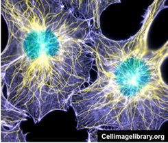
The cytoskeleton, pictured to the
right, consists of:
- Microfilaments (5-7 nm in
diameter)- composed of actin and regulate cell shape,
contractility, cell motility, and cytokinesis.
- Microtubules (24 nm in
diameter)- composed of tubulin dimers and contribute to cell
polarity and provide tracks for organelle and chromosome
movement.
- Intermediate filaments
have diameters between those of microfilaments and microtubules
(10-15 nm)- composed of a more diverse group of proteins and
provide strength and support to cells.
- Immunocytology to detect
intermediate filament proteins is used clinically to aid in
cancer diagnosis and treatment planning.Different cell types
express unique intermediate filament proteins (e.g.
epithelial cells express keratin intermediate filament
proteins, some neural cells express glial fibrillary acidic
protein, and muscle cells express desmin intermediate
filament proteins). Staining to detect
 these filaments can
help pathologists determine the origin of a tumor. these filaments can
help pathologists determine the origin of a tumor.
In routinely prepared H&E stained
specimens, the cytoskeleton contributes to the eosinophilia of
the cytoplasm. Immunocytology and fluorescence microscopy can be
used to reveal beautiful arrays of microtubules (yellow) and
microfilaments (purple).
The cytoskeleton, particularly
the microfilaments and microtubules, are highly dynamic
structures. Check out this live-imaging movie of a cell stained
with antibodies to detect
microtubules (green) and microfilaments (red). (Note, you
will need a Flash plug-in to view this movie.)
Notice the dynamic nature of the microfilaments and
microtubules, which can be seen here undergoing continuous
remodeling. In contrast, intermediate filaments are stable
structures that do not undergo continuous rearrangement.
How about the
nucleus? |