|
The nucleus, generally the largest
and most easily seen organelle in a cell, is filled with basophilic
chromatin. Chromatin consists of DNA associated with various
proteins and RNA. In routinely stained slides, nuclei can show
regions of euchromatin and heterochromatin in areas of high and low
gene activity respectively.
The nuclei of cells that are very actively synthesizing proteins,
will often contain abundant euchromatin and prominent nucleoli (the
site of rRNA synthesis and initial stages of ribosome assembly).
Examine the nuclei of neurons,
liver
cells, and cells of the pancreas,
and locate the euchromatin, heterochromatin, and nucleoli.
The
cells in these organs are particularly active in protein synthesis,
and their nuclei exhibit features that allow you to deduce this.

These
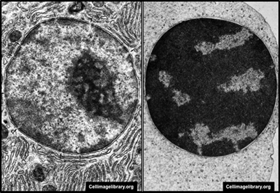
nuclear features are also easy to see in a TEM image of a pancreatic
cell (left panel). Contrast the pancreatic cell nucleus with the
nucleus of a terminally differentiating erythrocyte (right panel)
where extensive, inactive heterochromatin is present.
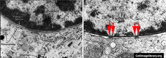
The nucleus is surrounded by the
nuclear envelope, which consists of two membranes:
- The outer is continuous with the
ER and the inner is associated with a layer of proteins (the
nuclear lamina) and heterochromatin.
- Passage of substances through
the envelope, in and out of the nucleus, is regulated by nuclear
pores.
Take a look at these TEM images
to see the nuclear envelope, lamina, and pores (arrows in right
image).

Cell division. |