Lysosomes and peroxisomes are somewhat similar in origin and appearance, but have
different functions.
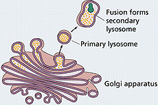
Lysosomes
contain numerous hydrolytic enzymes that allow them to degrade
endocytosed materials or cytoplasmic structures such as old or
damaged organelles. The process by which cells degrade their
unneeded components is called autophagy.
Peroxisomes contain oxidases and
participate in detoxification reactions and Beta-oxidation of long
chain fatty acids. Although they are abundant in metabolically
active cells such as hepatocytes and cells of the kidney
tubules, they are difficult to see in the LM without special
stains.
This material is sometimes
visible by LM in routinely stained cells, such as the
neurons and
liver
cells.
The remains of lysosomes
containing indigestible materials are called residual bodies or
lipofuscin granules. These are sometimes visible as aggregates
of brown staining material by LM in the cytoplasm of long-lived
cells, such as neurons and liver cells.
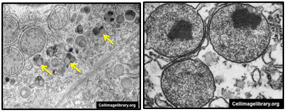
In TEM images, it can be seen
that lysosomes (arrows, left image) vary in size and electron density,
depending on what they are degrading. Peroxisomes (seen in the
TEM image below, right image) are more
uniform in size and electron density and sometimes contain a
small region of increased electron density corresponding to
their aggregated constituent enzymes.
 Lysosomes
contain numerous hydrolytic enzymes that allow them to degrade
endocytosed materials or cytoplasmic structures such as old or
damaged organelles. The process by which cells degrade their
unneeded components is called autophagy.
Lysosomes
contain numerous hydrolytic enzymes that allow them to degrade
endocytosed materials or cytoplasmic structures such as old or
damaged organelles. The process by which cells degrade their
unneeded components is called autophagy.