 Splenic
blood supply continued. Splenic
blood supply continued.
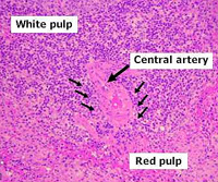
Identify central arterioles associated with the white pulp lymphoid
follicles (periarteriolar lymphoid sheathes or PALS), as well as the
marginal zone of these follicles.
In the red pulp identify the splenic
cords (of Billroth), seen at lower right, and the venous
sinuses. The latter are small in
this section of spleen,
but can be detected by their contents of red blood cells. Sheathed
vessels and sinuses are seen more readily in this
section of spleen, which is stained with a
special stain that also demonstrates elastic fibers in the trabeculae. Try to find a vascular sinus showing the unusual nature of the lining endothelial cells or stave cells.
Trace the highly unusual path of
blood in the spleen (through the arteries and into the venous
sinuses) and indicate how this pathway relates to the spleen's
function.
What is the functional
relationship between the venous sinuses and the surrounding red
pulp?
Next is the
digestive system. |