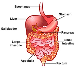
 The digestive system consists
of a long muscular tube, extending from mouth to anus, and a large
number of associated glands, which secrete substances that aid food
digestion and absorption of nutrients. The digestive tube is
generally made up of four structurally and functionally distinct
layers: the mucosa, submucosa, muscularis, and serosa
(or the adventitia, if not covered by serosa). The mucosa,
which is in contact with the luminal contents, is constructed to
resist abrasion and to perform secretory and absorptive functions.
The muscular layer, which serves to propel the food through the
tube, is attached to the mucosa by a dense connective tissue layer,
the submucosa. The serosa or adventitia carries blood vessels and
nerves to the wall of the digestive tube. The digestive system consists
of a long muscular tube, extending from mouth to anus, and a large
number of associated glands, which secrete substances that aid food
digestion and absorption of nutrients. The digestive tube is
generally made up of four structurally and functionally distinct
layers: the mucosa, submucosa, muscularis, and serosa
(or the adventitia, if not covered by serosa). The mucosa,
which is in contact with the luminal contents, is constructed to
resist abrasion and to perform secretory and absorptive functions.
The muscular layer, which serves to propel the food through the
tube, is attached to the mucosa by a dense connective tissue layer,
the submucosa. The serosa or adventitia carries blood vessels and
nerves to the wall of the digestive tube.
The large number of glands associated
with the digestive tract range from unicellular and small
tubuloalveolar glands residing in the mucosa to large organs such as
the pancreas and liver. Although their secretions can be quite
dissimilar, they all function to promote the digestive process by
imparting enzymes or mucus into the ingested food or by regulating
motility of the tract to propel the ingested food distally.
The learning objectives for this
unit are:
- Identify the histologic and
cellular features of teeth and their supportive structures.
- Identify the histoarchitecture
of the tongue, including the gustatory and support cells in
taste buds, and the functional significance of these structures.
- Compare and contrast the
secretory functions and relative quantities of serous and mucous
cells in parotid, submandibular, and sublingual glands.
- Identify the histological layers
and unique cellular characteristics associated with the oral
cavity, esophagus, stomach, duodenum, jejunum, ileum, appendix,
and colon (mucosa, submucosa, muscularis proper, and serosa/adventitia)
and their functions.
Let's start with the
oral cavity. |