|
The thymus is the primary lymphoid organ
where immature T cells, thymocytes, mature into the specific
subpopulations of T cells (e.g. helper or cytotoxic T cells) and
gain central tolerance. The development of central tolerance
is crucial so that these cells do not become activated by self-antigens.
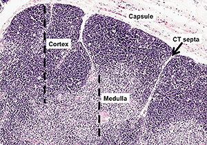 Examine this
section of thymus Examine this
section of thymus and the image
at the right, noting the capsule, connective tissue septa, and
organization of lobes into a basophilic cortex and a paler staining
medulla. and the image
at the right, noting the capsule, connective tissue septa, and
organization of lobes into a basophilic cortex and a paler staining
medulla.
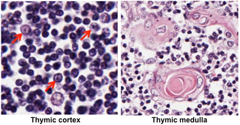
Identify the thymic epithelial cells
(also called epithelial-reticular cells or epitheliocytes), which
make up the framework of the thymus. In the cortex as seen below,
these cells are larger than the lymphocytes, and their nuclei
contain prominent nucleoli. In the medulla, identify the thymic
corpuscles or Hassall corpuscles, which are large collections of
thymic epithelial cells. Thymic epithelial cells function to 1) form
a blood-thymus barrier to shield developing thymocytes from exposure
to antigens, 2) compartmentalize the thymus to sequester immature
and mature thymocytes, in the cortex and medulla, respectively, 3)
provide a structural and supportive framework for the developing
thymocytes (the thymus lacks reticular fibers), and 4) present self
and foreign antigens to developing thymocytes during positive and
negative selection.
Last, examine a section of
involuted
thymus, and
note the accumulation of adipocytes and depletion of lymphocytes.
Next let's look at
lymph nodes. |