|
Before we look at the organs of the
lymphoid system, let’s first review the principal cells of the
lymphoid system, the lymphocytes. Lymphocytes are produced in
primary lymphoid organs (bone marrow and thymus), and with the help
of antigen presenting cells, such as macrophages and dendritic
cells, they are activated to respond to specific antigens in
secondary lymphoid organs (lymphatic nodules, lymph nodes, and
spleen). There are two main types of lymphocytes, B and T
lymphocytes.
- B lymphocytes or simply B
cells, possess B cell receptors (BCRs) on their cell surfaces
that bind antigens. In response to antigen binding, B cells
proliferate and differentiate to give rise to plasma cells that
produce a single type of antibody that is specific for that
antigen. During this differentiation process, some of these
activated B cells become memory B cells. These long-lived cells
are capable of undergoing rapid activation to produce plasma
cells when the body encounters that particular antigen again.
- T lymphocytes express T
cell receptors (TCRs) on their cell surfaces and are found as
several different subtypes in the body, including Helper T cells
(CD4+), Cytotoxic T cells (CD8+), and Regulatory T cells. Helper
T cells assist in activation of other lymphocytes and also form
populations of memory cells that can be reactivated in response
to future antigen exposure. Cytotoxic T cells are specialized
for cell-mediated killing of virus-infected and foreign cells.
The activation of cytotoxic T cells also yields populations of
memory T cells. Finally, Regulatory T cells maintain tolerance
of self-antigens and help keep immune responses in check.
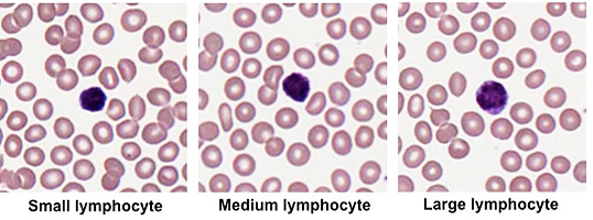
- Now examine this
blood smear slide, focusing
your attention on the structure of the lymphocytes. Lymphocytes
range in size of 6-15 microns in diameter and are sometimes
described as small, medium, and large lymphocytes (see image
below). They have a large, round centrally located nucleus with
a thin rim of pale blue cytoplasm. It is not possible to
distinguish the different types of lymphocytes (e.g. B vs. T
cells) in a histological specimen without the help of immunocytology that allows for visualization of these
populations based on the detection of specific cell surface
proteins.
Next let's look at
the thymus. |