|
The lens is a transparent biconvex
disc suspended behind the iris by the ciliary zonules that extend
from the ciliary body. Contraction and relaxation of the ciliary
muscles changes the shape of the lens, allowing for visual
accommodation (focus on near and far objects). The lens surface is
surrounded by a proteoglycan and type IV collagen-rich lens
capsule to which the ciliary zonules attach. Below the lens
capsule on the anterior surface is the lens epithelium, a
simple cuboidal epithelium. At the posterior edge near the lens
equator, the lens epithelium proliferates and terminally
differentiates, giving rise to lens fibers that lack
organelles and are filled with crystallins. The tightly packed lens
fibers provide for transparency of the lens.
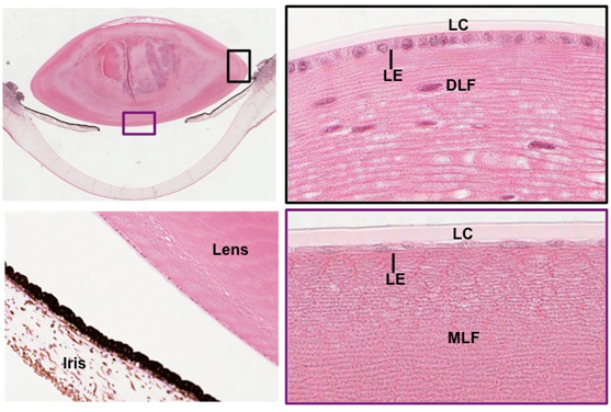
- Examine the lens in the images below and
this slide. Identify
the capsule (LC) and simple cuboidal lens epithelium (LE),
differentiating nucleated lens fibers (DLF), and mature
non-nucleated lens fibers (MLF).

Clinical note: Oxidative changes in
the lens fibers are common as one ages and can lead to opacity of
lens tissue called a cataract, which eventually produces blurred
vision. In surgery for cataracts, such lenses are broken up and
aspirated out through a slit in the upper cornea, leaving the zonule
and thick posterior capsule of the lens in place to hold an
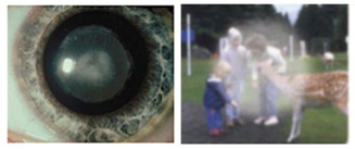
implanted plastic lens. The two images to the right show what a
cataract lens looks like and what a person sees with such a lens.


- Next, examine the images at the
right and the iris on this slide,
noting the continuity with the ciliary body. Identify the
heavily pigmented epithelium (PE), the stroma (S), the
sphincter pupillae muscle (SPM, a circular bundle of smooth muscle near
the pupil), and the dilator pupillae muscle (DPM, a very thin
layer consisting of contractile processes of myoepithelial
cells). Identify the anterior (AC) and posterior (PC) chambers,
meeting at the pupil.
Now for the
cornea. |