|
In the images below and on
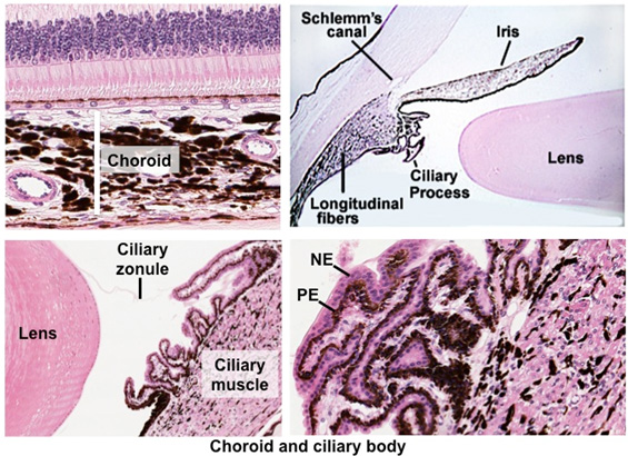
this slide, identify
the heavily pigmented choroid layer surrounding the retina. Follow
the choroid anteriorly and locate the ciliary body. Identify smooth
muscle in the ciliary body, projecting ciliary processes, and
remnants of the ciliary zonule or suspensory ligaments. Note the
bilayered epithelium, inner pigmented epithelium (PE) and outer
non-pigmented epithelium (NP), lining the ciliary processes. This
epithelium secretes aqueous humor and attaches to the lens via the
zonule fibers. At the limbus, near where the ciliary body and iris
join the cornea, identify the endothelium-lined spaces of the canal
of Schlemm, which drains the aqueous humor flowing through the
anterior chamber.
  Clinical
note: Glaucoma results from the decreased outflow of aqueous
humor through the trabecular meshwork and the canal of Schlemm. This
elevates intraocular pressure, which can damage peripheral areas of
the retina and cause progressive loss of vision. Various medications
can lower the pressure and laser surgery can be used to improve
drainage through the trabecular meshwork. To the right is how an
individual with glaucoma sees the world. Clinical
note: Glaucoma results from the decreased outflow of aqueous
humor through the trabecular meshwork and the canal of Schlemm. This
elevates intraocular pressure, which can damage peripheral areas of
the retina and cause progressive loss of vision. Various medications
can lower the pressure and laser surgery can be used to improve
drainage through the trabecular meshwork. To the right is how an
individual with glaucoma sees the world.
The lens is next. |