|
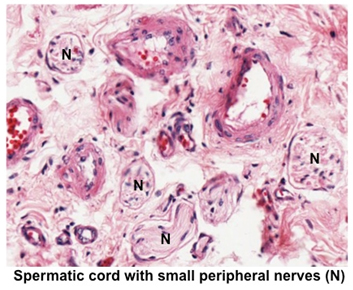 In the next few slides we’ll study
the organization of peripheral nerves. Examine sections of
small autonomic ganglia
and small peripheral
nerves in loose CT and in this
section of the
spermatic cord.
Identify the nerve fibers, Schwann cells, and surrounding CT layer. In the next few slides we’ll study
the organization of peripheral nerves. Examine sections of
small autonomic ganglia
and small peripheral
nerves in loose CT and in this
section of the
spermatic cord.
Identify the nerve fibers, Schwann cells, and surrounding CT layer.
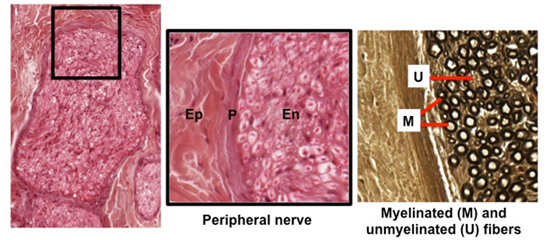
Examine the images below and these two sections of peripheral
nerve (sample 1 ,
sample 2). Sample
2 has been prepared with osmium, which stains myelin black. The perineurium, along with the nature of the capillaries in the
endoneurium, make up a “blood-nerve barrier” that helps protect the
myelin sheaths of peripheral nerves from viral or immunological
agents. In these specimens identify: ,
sample 2). Sample
2 has been prepared with osmium, which stains myelin black. The perineurium, along with the nature of the capillaries in the
endoneurium, make up a “blood-nerve barrier” that helps protect the
myelin sheaths of peripheral nerves from viral or immunological
agents. In these specimens identify:
- Epineurium (Ep)- thick dense
irregular CT surrounding several nerves
- Perineurium (P)- CT layer
surrounding a group of nerve fibers
- Endoneurium (En)- very thin CT
layer surrounding Schwann cells in nerves
- Myelinated fibers
- Non-myelinated fibers
Next are
synapses and sensory receptors. |