| Skeletal or Striated Muscle
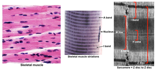
Skeletal muscle tissue is distinctive
due to its long, tube-shaped multinucleated fibers (cells) with
visible cross-striations. The striations are the result of highly
ordered actin and myosin filaments within sarcomeres, the
basic contractile units of muscle fibers (see images below). Sliding
of the myosin thick filaments along the actin thin filaments within
the sarcomeres leads to contraction of striated muscle fibers.
Review the organization of the bands in the sarcomeres.
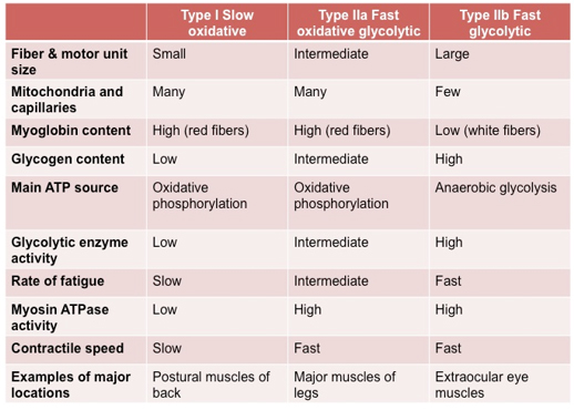
Skeletal muscle contains three main
fiber types that are distinguished based on several properties, such
as size of the fibers, their contractile rates, and the main
pathways used to generate ATP. Review the table below describing the
three types of fibers: type I slow oxidative, type IIa fast
oxidative glycolytic, and type IIb fast glycolytic fibers.
 Examine skeletal muscle in the images
below and in these slides of the larynx (sample
1 and
sample 2) and
tongue Examine skeletal muscle in the images
below and in these slides of the larynx (sample
1 and
sample 2) and
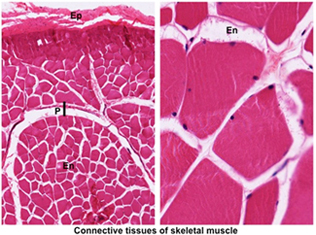
tongue using low power. Study its organization into fascicles,
and identify the connective tissues of muscle: using low power. Study its organization into fascicles,
and identify the connective tissues of muscle:
- Epimysium (Ep), a
dense irregular CT that surrounds the entire muscle
- Perimysium (P), a
thinner CT layer that bundles muscle fibers into a fascicle
- Endomysium (En), a fine CT layer that surrounds each muscle fiber
At higher power, examine muscle fibers, and identify endomysium
and blood vessels of various sizes. Examine the transversely cut
skeletal muscle fibers in the H&E stained
specimen of larynx
again. Try to
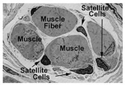
distinguish individual myofibrils within the muscle fibers. Note the
position of the nuclei at the periphery of the muscle fibers. Nuclei
of satellite cells (a limited population of progenitor cells capable
of giving rise to new muscle fibers after muscle damage due to
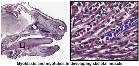
disease or injury) are also located here. Examine the images below
and developing skeletal muscle of the
fetal tongue and face
to see myoblasts (that give rise to satellite cells) and developing
muscle fibers (myotubes).
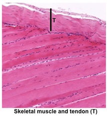
Finally, examine the dense regular
connective tissue of tendon
slide and compare its features to those of
skeletal muscle. Note the differences in the location and shape of
the nuclei and absence of striations in the tendon (seen in the
image at the right).
 Clinical note: Exercise or increased use of
specific muscles produce hypertrophy or increased fiber size, while
disuse results in muscle atrophy. Muscular dystrophies, a group of
hereditary diseases, are characterized by muscle weakness and
atrophy. Recent research has shown that in one form of this disease, Duchenne muscular dystrophy, abnormal function of satellite cells
leads to impaired regeneration. Clinical note: Exercise or increased use of
specific muscles produce hypertrophy or increased fiber size, while
disuse results in muscle atrophy. Muscular dystrophies, a group of
hereditary diseases, are characterized by muscle weakness and
atrophy. Recent research has shown that in one form of this disease, Duchenne muscular dystrophy, abnormal function of satellite cells
leads to impaired regeneration.
Next is a little
more about muscle contraction. |