|
 Movement of body parts depends
primarily on muscle tissue, which is highly specialized for
contraction. The importance of this tissue is emphasized by the fact
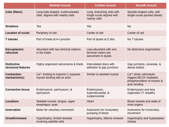
that almost half the body's mass consists of muscle. There are three
types of muscle fibers. Compare and contrast their properties in the
table below. Movement of body parts depends
primarily on muscle tissue, which is highly specialized for
contraction. The importance of this tissue is emphasized by the fact
that almost half the body's mass consists of muscle. There are three
types of muscle fibers. Compare and contrast their properties in the
table below.
- Skeletal muscle that is
primarily involved in movement of bones (voluntary)
- Cardiac muscle that enables the
heart to beat so that blood can be circulated (involuntary)
- Smooth muscle (visceral muscle)
that provides tone and movement of hollow tubes and organs such
as the intestines and uterus (involuntary)
The learning objectives for this
unit are:
- Identify the key structural
features of the three muscle types (skeletal, cardiac, and
smooth) by light and electron microscopy, and explain how
each is organized to form a contractile tissue that performs
specific types of work.
- Recognize the key features
of neuromuscular junctions by electron microscopy and
discuss their functional significance.
- Compare and contrast the
arrangement of actin and myosin filaments in skeletal,
cardiac, and smooth muscle in electron micrographs.
- Compare and contrast the
arrangement and functions of transverse tubules,
sarcoplasmic reticulum, mitochondria, and contractile
filaments in electron micrographs of skeletal and cardiac
muscle.
- Identify the structural and
functional attributes of connective tissues associated with
muscle and the myotendinous junction.
Let's begin with
skeletal or striated muscle. |