|
Connective Tissue Proper
Connective tissue proper varies
in its density and arrangement of collagen bundles and generally
can be described as either “loose” or “dense.” Again, these are
relative terms, and the lines separating these classifications
are not distinct but graded.
Loose Connective Tissue Proper
Relative to dense CT, loose CT
is typically highly cellular and contains sparse, thin, and
loosely arranged fibers and abundant ground substance. Loose CT
can further be described as areolar or reticular.
Loose (areolar) connective tissue
Loose (areolar) CT is
characterized by relatively loosely arranged collagen and
elastic fibers. It is highly cellular, containing fibroblasts,
immune cells (e.g., mast cells, macrophages, T cells), and
endothelial cells (capillaries). In some cases, you’ll find
adipocytes and greater amounts of unstained regions due to
extraction of ground substance during tissue processing. It is
often found directly underlying epithelia that cover body
surfaces or line internal surfaces. In mucosal epithelium
(respiratory system, alimentary canal, & genitourinary tract) it
is called lamina propria.
 You can also find it associated with
glandular epithelium and surrounding small blood vessels. You can also find it associated with
glandular epithelium and surrounding small blood vessels.
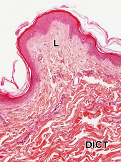
The papillary dermis (connective
tissue right underneath skin epithelium; see image to the right) has
small collagen bundles that are pretty tightly packed but is still
considered loose or areolar connective tissue (L). As you examine
this slide of human thin
skin, note
that the distinction between loose CT (L) and dense irregular
connective tissue (DICT) is clearer when they are viewed in
juxtaposition. note
that the distinction between loose CT (L) and dense irregular
connective tissue (DICT) is clearer when they are viewed in
juxtaposition.
Examine the connective tissue, both loose connective tissue (also
known as lamina propria in the colon) and dense irregular connective
tissue, and compare it to the appearance of smooth muscle on this
slide of the colon
. Examine the CT in dermis of the skin in the
following examples: thin
skin 1, thin skin 2,
thick skin 1, and
thick skin 2, which
have different stains, noting again the different densities of CT.

Clinical note: Following injury
to any tissue or organ, connective tissue proper is the usual site
of the inflammatory response, a process that involves all the cells
of the tissue (cells of CT are discussed later). The events of wound
healing mainly involve cells and fibers of the connective tissue and
the formation of scars involves excessive or otherwise abnormal
production of new fibroblasts and collagen during this process.
Reticular Tissue
Loose reticular CT
contains a network of reticular fibers of type III collagen (also
called “reticulin”). These fibers are produced by specialized
fibroblasts (a.k.a reticular cells) and form an elaborate network
through which interstitial fluid or lymph and wandering blood cells
pass continuously. Reticulin fibers stain black with silver stains,
as seen in this oval
lymph node .
Also note the appearance of the collagen bundles in the CT
surrounding the lymph node, comparing it with that of the stains
examined earlier.
Now let's examine
dense CT proper, both dense irregular
and dense regular CT. |