|
The paired seminal vesicles are
tortuous tubes surrounded by a connective tissue capsule. Their
mucosa is highly folded, making it falsely appear as if these glands
have multiple lumens. The lining epithelium is simple cuboidal/columnar
or pseudostratified columnar epithelium. Underlying
the mucosa is a fibroelastic lamina propria and two layers of smooth
muscle. The seminal vesicles secrete a fluid that is rich in
fructose, prostaglandins, and fibrinogen, and constitutes ~70% of
the fluid component of semen.
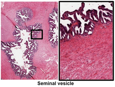
- Examine the images at the right
and a section of the
seminal vesicle. Identify columnar/cuboidal epithelium (it
may appear pseudostratified) in the thin, highly folded mucosa
and the layers of smooth muscle surrounding the secretory
components.
The prostate gland consists of
tubuloacinar glands lined by a simple or pseudostratified columnar
epithelium surrounded by a fibromuscular stroma. The ejaculatory
duct, carrying sperm in seminal fluid, empties into the prostatic
urethra that runs through the center of the gland. The prostate
gland secretes a fluid containing enzymes, glycoproteins, and
prostaglandins that is added to the semen during ejaculation. The
lumens of the prostate gland sometimes contain small concretions, or
corpora amylacea, which are helpful in identifying the
prostate gland, but have no known functional or clinical
significance.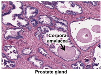
- Examine the image at the right
and this trichrome-stained
section of prostate gland.
Identify the tall epithelial cells with their lightly stained
and "foamy" appearing apical cytoplasm. In the stroma, notice
the smooth muscle fibers (red) mixed with dense
connective tissue (blue). In the lumens, identify the calcified, proteinaceous concretions, corpora amylacea.
- The association between
prostate and urethra
 can be seen in this specimen, which shows the region near
the urethra. The urethra has been split and appears to be on
the edge of the section. Notice the difference between the
glandular tissue near the urethra and the glands throughout
most of the organ further from the urethra.
can be seen in this specimen, which shows the region near
the urethra. The urethra has been split and appears to be on
the edge of the section. Notice the difference between the
glandular tissue near the urethra and the glands throughout
most of the organ further from the urethra.
Clinical note: Prostate
glands provide urologists with plenty of work by being prone
to three very common problems. (1) They are the sites of
chronic, low-grade bacterial infections. (2) In older persons
the secretory epithelium very frequently undergoes benign
hyperplasia. This tissue overgrowth constricts the urethra,
causing problems with urination. (3) Adenocarcinoma of the
prostate epithelium is a very common form of cancer.
The penis and
urethra are next. |