On this
section of epididymis ,
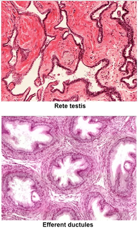
and in the image at the right, identify the efferent ducts with
their characteristic saw tooth or scalloped appearance due to the
presence of tall columnar ciliated epithelial cells interspersed
with simple cuboidal cells. The efferent ducts absorb most of the
fluid produced by the seminiferous tubules and convey the sperm into
the epididymis via contraction of the smooth muscle surrounding the
ducts. ,
and in the image at the right, identify the efferent ducts with
their characteristic saw tooth or scalloped appearance due to the
presence of tall columnar ciliated epithelial cells interspersed
with simple cuboidal cells. The efferent ducts absorb most of the
fluid produced by the seminiferous tubules and convey the sperm into
the epididymis via contraction of the smooth muscle surrounding the
ducts.
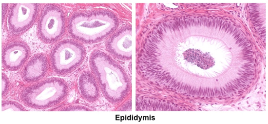
The
epididymis is a long convoluted duct with smooth muscle in its wall.
Examine the images below and the beginning or
head of the epididymis
adjacent to the efferent ductules and the tail of the epididymis
(leading to the vas deferens). Identify the tall columnar, pseudostratified epithelium with
stereocilia and the stored
spermatozoa in the lumen. The epididymis functions to promote
maturation of the sperm, providing factors that decapacitate them
(prevent their fertilizing ability) and allow them to become fully
motile. The epididymis also stores the spermatozoa and propels them
into the ductus deferens during ejaculation.
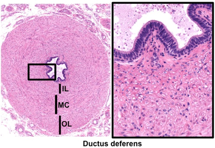
The ductus deferens or vas
deferens is a muscular tube that leads from the epididymis to
the prostate gland and functions to rapidly propel sperm during
ejaculation.
- Examine the images below and
these specimens of ductus deferens (sample
1, sample 2).
At higher magnification, identify the pseudostratified columnar
epithelium with sparse stereocilia, fibroelastic lamina propria,
and three thick layers of smooth muscle (organized as inner
longitudinal (IL), middle circular (MC), and outer longitudinal
(OL) layers) that allow for rapid, peristaltic expulsion of
sperm from the epididymis. In sample 2, note the arteries,
veins, and nerves embedded in loose connective tissue that
travel with the ductus deferens in the spermatic cord.
Now for the prostate and seminal vesicle. |