|
The Uterus is the site of
implantation and growth of an embryo. The endometrium undergoes marked
changes during the hormonal cycle, resulting in distinctive
histological features during the menstrual, proliferative, and secretory
phases. 
Examine two sections of uterus (sample
1 - trichrome stain, and
sample
2 - H&E) and identify the
- Endometrium,
- Myometrium,
- Perimetrium with serosa, if
present, and
- The attached connective tissue, if
present.
Examine the endometrial surface and
note the simple columnar epithelium, with many ciliated cells.
Throughout the endometrium, identify endometrial glands and vascular
sinuses in the stroma. You may be able to distinguish the basal
layer and functional layer of the endometrium, but this is difficult
on sample 2. In the myometrium, observe the interlacing bundles of
smooth muscle fibers.
Clinical note: Endometriosis, results when sloughed endometrial tissue
is refluxed up the uterine tubes and surviving cells begin to grow
on the surface of the ovary, mesenteries, or other organs in the
peritoneal cavity. Cycles of growth and loss of this displaced
endometrial tissue occur under the influence of estrogen and
progesterone, which can eventually result in inflammation, pain, and
scarring of the affected organs. Untreated, endometriosis can lead
to infertility for several reasons.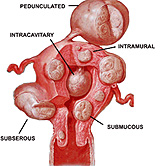
Clinical note: Smooth muscle
cells of the uterus commonly give rise to benign tumors called
leiomyomas or fibroids. They grow slowly and often attain diameters
of several centimeters, but usually do not produce major medical
problems.
Now let's look at the
stages of endometrial development. |