|
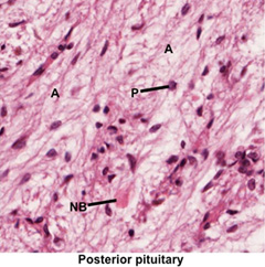
The posterior pituitary or
neurohypophysis is a neurosecretory gland containing axons of
secretory neurons whose cell bodies reside in the supraoptic and
paraventricular nuclei. These neurons produce oxytocin and
antidiuretic hormone (ADH or vasopressin), which are released at the
axon termini that are closely associated with capillaries.
Occasionally, the axon termini enlarge due to accumulation of
secretory vesicles and are visible as neurosecretory bodies
(NB, Herring bodies).
 Oxytocin
stimulates myoepithelial cell contraction during lactation and
smooth muscle contraction in the myometrium of the uterus.
Antidiuretic hormone acts on the kidney collecting ducts to increase
the absorption of water. Oxytocin
stimulates myoepithelial cell contraction during lactation and
smooth muscle contraction in the myometrium of the uterus.
Antidiuretic hormone acts on the kidney collecting ducts to increase
the absorption of water.
- Examine the image at the right
and these slides containing the posterior pituitary (sample
1, sample 2).
Identify capillaries, non-myelinated axons of secretory
neurons (A), and their supporting cells called pituicytes
(P).
Clinical note: Diabetes insipidus
is a disorder in which the neurohypophysis fails to secrete
antidiuretic hormone in response to normal stimuli. The disorder
involves excessive thirst, water intake, and urination and can
be treated by injection of vasopressin or similar drugs.
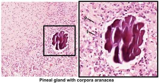
The pineal gland or epiphysis cerebri is a small neurosecretory
organ in the brain containing secretory neurons (pinealocytes),
interstitial glial cells, and axons that communicate with other
parts of the brain. The pineal gland functions to regulate the
body’s circadian rhythms by relating light intensity and
duration to hormone secretion. Melatonin, the major hormone
secreted by the pinealocytes, regulates the activity of the
hypothalamus, pituitary, and other endocrine organs. The
interstitial glial cells support the pinealocytes and axons
within the gland. Pia mater surrounds the gland, and connective
tissue septa extend from the pia mater into the gland, dividing
it into irregular lobules. A distinctive feature of the pineal
gland is the presence of corpora aranacea (brain sand),
concretions of mineralized extracellular proteins of unknown
function. Brain sand has clinical relevance because radiologists
find that it is a good marker of the brain’s midline.
Examine the
pineal gland in the images below and on these two slides (sample
1, sample 2). Pineal
tissue can be identified from surrounding brain tissue by the
presence of brain sand. Identify the abundant vasculature, small
clusters of pinealocytes (P), with scant, poorly stained
cytoplasm and ovoid euchromatic nuclei. Distinguish the
interstitial glial cells (I), which are smaller than the pinealocytes and typically have elongated, darker staining
nuclei. Note the fibrous material surrounding the cells is
primarily neuropil.
Next is the
thyroid. |