- Study the images below and this
specimen of
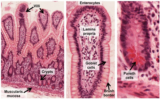
ileum at higher magnification. In the villi, identify the
simple columnar absorptive cells or enterocytes with
their brush border (microvilli or striated border),
terminal web, and goblet cells.
In the lamina propria, note the abundant lymphocytes, and
locate the central lacteal. The central lacteals are
lymphatics that function in immune response and transport of
chylomicrons. Also in the lamina propria, note the presence
of capillaries for transfer of absorbed molecules into the
blood. Identify the crypts containing mitotic cells that
renew the lining epithelium and Paneth cells with their
distinctive, eosinophilic secretory granules (containing
antimicrobial substances, e.g. lysozyme and defensins). Last,
locate the muscularis mucosa.

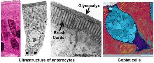
- Next, in the images below,
review the ultrastructural features of the enterocytes,
including the brush or striated border (microvilli) with a
glycocalyx that contains enzymes involved in digestion. Study
the colorized TEM image of a goblet cell among the
enterocytes. Note the unique shape of these cells and the
abundant mucin secretory granules (colored blue) within the
apical cytoplasm.


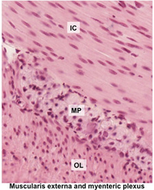
- Finally, in the image at the
right and the
specimen of duodenum on this slide, examine the
muscularis externa, identifying the inner circular (IC) and
outer longitudinal (OL) layers of smooth muscle. In between the
layers, locate the myenteric (Auerbach) plexus.
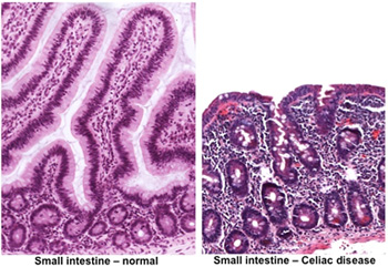
 Clinical note: A disorder
called celiac disease (celiac sprue, gluten-induced enteropathy) is
characterized by intolerance to gluten (a protein in many grains).
It results in loss of enterocyte microvilli, flattening of the villi,
and subsequent malabsorption of nutrients. Regeneration of these
structures in the small bowel and return of normal nutrient
absorption occur after a few weeks on a diet lacking gluten. Clinical note: A disorder
called celiac disease (celiac sprue, gluten-induced enteropathy) is
characterized by intolerance to gluten (a protein in many grains).
It results in loss of enterocyte microvilli, flattening of the villi,
and subsequent malabsorption of nutrients. Regeneration of these
structures in the small bowel and return of normal nutrient
absorption occur after a few weeks on a diet lacking gluten.
Next is large
intestine. |