|
 The small intestine is the
site where digestion is completed, using enzymes from the pancreas
and bile, and where products of digestion are absorbed. The small
intestine possesses several features that increase the surface area
for digestion and absorption, including its long length, the
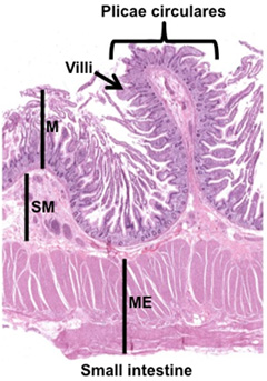
presence of plicae circulares, numerous villi, and the simple
columnar epithelium with microvilli (brush border). As seen in the
image at the right, the four general layers of the GI tract (mucosa
(M), submucosa (SM), muscularis externa (ME), and serosa/adventitia)
are present in the small intestine. The three regions of the small
intestine, the duodenum, jejunum, and ileum, are
similar histologically with a few distinguishing features. The small intestine is the
site where digestion is completed, using enzymes from the pancreas
and bile, and where products of digestion are absorbed. The small
intestine possesses several features that increase the surface area
for digestion and absorption, including its long length, the
presence of plicae circulares, numerous villi, and the simple
columnar epithelium with microvilli (brush border). As seen in the
image at the right, the four general layers of the GI tract (mucosa
(M), submucosa (SM), muscularis externa (ME), and serosa/adventitia)
are present in the small intestine. The three regions of the small
intestine, the duodenum, jejunum, and ileum, are
similar histologically with a few distinguishing features.
- Examine these two sections of
the duodenum (sample 1,
sample 2).
Identify the major layers of the small intestine and observe the
plicae circulares with tightly packed villi formed as
projections of the mucosa layer. Study the villi, crypts (of Lieberkuhn), muscularis mucosa, submucosal Brunner’s glands
(BG), inner circular and outer longitudinal layers of the muscularis externa, and adventitia.
- Examine two sections of
jejunum (sample
1
 , sample 2)
and ileum (sample 1,
sample 2).
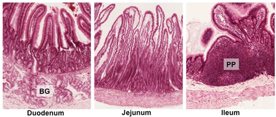
For a comparison of all three regions, examine this
small
intestine composite specimen.
Compare and contrast the features of each region in the slides
and images below. Note that the duodenum contains Brunner’s
glands (BG) and the ileum contains Peyer’s patches (PP), which
are distinguishing features. , sample 2)
and ileum (sample 1,
sample 2).
For a comparison of all three regions, examine this
small
intestine composite specimen.
Compare and contrast the features of each region in the slides
and images below. Note that the duodenum contains Brunner’s
glands (BG) and the ileum contains Peyer’s patches (PP), which
are distinguishing features.
Now let's
take a closer look at the small intestines. |