|
 The stomach is the site where
food is converted to a thick fluid called chyme that allows for
efficient enzymatic digestion of macromolecules. The wall of the
stomach contains gastric folds or rugae that expand when the stomach
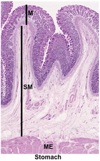
is full. As seen in the image at the right, the stomach contains the
typical layers seen in the GI tract, including the mucosa (M,
lining epithelium and glands, lamina propria, and muscularis
mucosa), submucosa (SM), muscularis externa (ME,
containing three layers, oblique, circular, and longitudinal layers
of smooth muscle), and a serosa. The stomach is the site where
food is converted to a thick fluid called chyme that allows for
efficient enzymatic digestion of macromolecules. The wall of the
stomach contains gastric folds or rugae that expand when the stomach
is full. As seen in the image at the right, the stomach contains the
typical layers seen in the GI tract, including the mucosa (M,
lining epithelium and glands, lamina propria, and muscularis
mucosa), submucosa (SM), muscularis externa (ME,
containing three layers, oblique, circular, and longitudinal layers
of smooth muscle), and a serosa.
Histologically, three regions are
distinguishable in the stomach: the cardia, fundus/body,
and pylorus. The cardia is the transitional region between
the esophagus and body of the stomach. The pylorus is the narrow
region that connects the stomach to the duodenum. The cardia and
pylorus contain branched coiled tubular glands that secrete mucus.
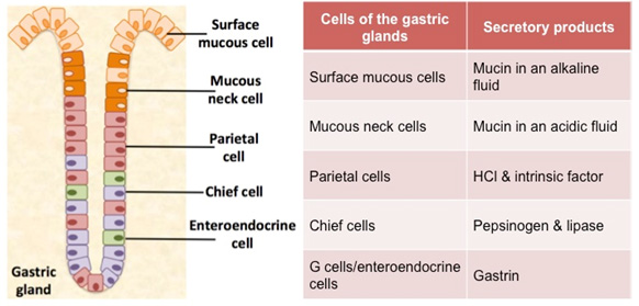
In contrast, the mucosa of the fundus/body contains long, branched
tubular gastric glands with several cell types that secrete
substances that aid in digestion. See the diagram and table below to
review the cells and secretions of the gastric glands.

- Examine the images below and
this section of the
stomach fundus and body
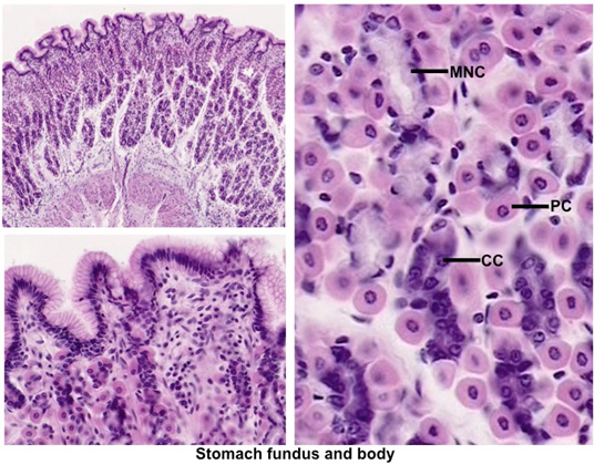
 . Identify the layers and sublayers,
noting that the simple columnar epithelium invaginates into
gastric pits, which are tightly packed together. These lead into
the gastric glands, which extend down to the thin muscularis
mucosa. Cells of the lamina propria are seen scattered loosely
around the gastric pits. . Identify the layers and sublayers,
noting that the simple columnar epithelium invaginates into
gastric pits, which are tightly packed together. These lead into
the gastric glands, which extend down to the thin muscularis
mucosa. Cells of the lamina propria are seen scattered loosely
around the gastric pits.
- On the same slide of stomach,
find a region of mucosa in which the gastric glands are cut
longitudinally. Starting at the surface, identify the
mucus-secreting cells (called surface and neck cells depending
on their proximity to the surface lining the stomach cavity),
the large eosinophilic parietal cells, and the smaller,
more basophilic peptic chief cells. Enteroendocrine
cells are also present (e.g. G cells that secrete gastrin), but
these cells are especially difficult to identify without special
stains.

- Examine the images below and
these slides of the
mucosa of the
cardia and
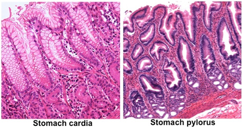
mucosa of the pyloric stomach. Note that the pyloric glands,
unlike the gastric glands found in the fundus and body of the
stomach, contain mainly mucus-secreting cells with few parietal
and gastrin secreting cells.

On to the
small intestine. |