|
 The esophagus is a muscular
tube that conducts food from the oral cavity to the stomach. Review
the general organization of the gastrointestinal tract, noting
especially the four major layers, which are clearly seen in the
esophagus: The esophagus is a muscular
tube that conducts food from the oral cavity to the stomach. Review
the general organization of the gastrointestinal tract, noting
especially the four major layers, which are clearly seen in the
esophagus:
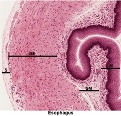
- Mucosa (M) - epithelia,
lamina propria, and muscularis mucosa
- Submucosa (SM) - dense
irregular connective tissue containing blood vessels, lymphatics,
and nerves
- Muscularis externa (ME) -
two thick layers of smooth muscle for peristalsis
- Adventitia or serosa (A or S)
- outer connective tissue or epithelial covering
 Examine the images below and
transverse sections of the esophagus (sample
1, sample 2).
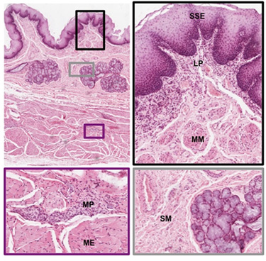
Identify the various layers of the wall, including: Examine the images below and
transverse sections of the esophagus (sample
1, sample 2).
Identify the various layers of the wall, including:
- Stratified squamous
epithelium (SSE)
- Lamina propria (LP)
- Muscularis mucosa (MM)
- Submucosa (SM) CT with small
mucous glands and lymphoid nodules
- Myenteric plexus (MP) with
autonomic ganglia and nerves
- Muscularis externa (ME)
(striated muscle of upper 1/3, mixed striated and smooth muscle
of middle 1/3, and smooth muscle of lower 1/3)
- Adventitia or serosa
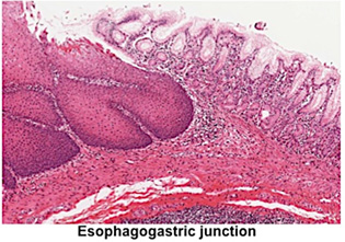
Examine these two sections of the
esophagogastric junction (sample
1, sample 2).
Note the abrupt, change in the lining epithelium from stratified
squamous to simple columnar epithelium.
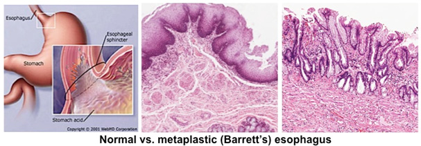
Clinical note: If the
muscularis of the lower esophagus fails to maintain the state of
partial contraction that normally prevents reflux of the gastric
contents upward, heartburn results. Since the esophageal mucosa
lacks a thick blanket of protective mucus, acidic gastric juices
irritate it, causing painful esophagitis. If chronic, this is called
gastro-esophageal reflux disorder (GERD) and can lead to metaplasia
of stratified squamous epithelium into a simple columnar, mucous
epithelium. (Image courtesy of WebMD)
Now to the stomach. |