|
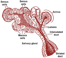 The oral cavity contains three pairs
of major salivary glands, the parotid, submandibular,
and sublingual, and many minor salivary glands within the
submucosa of the oral cavity. Salivary glands are compound acinar
glands surrounded by a connective tissue capsule (review their
structure in the schematic at the right). Connective tissue septa
extend into the gland dividing it into lobes and lobules. Secretions
from the acini are released into the acinar lumens and then travel
through intercalated ducts, striated ducts, and then into
excretory ducts that open into the oral cavity. The parotid
gland produces serous secretions, whereas the sublingual and
submandibular produce mixed seromucous secretions. The submandibular
and sublingual glands can be distinguished because the sublingual
glands contain more mucous acini. The minor salivary glands mainly
secrete mucus. The oral cavity contains three pairs
of major salivary glands, the parotid, submandibular,
and sublingual, and many minor salivary glands within the
submucosa of the oral cavity. Salivary glands are compound acinar
glands surrounded by a connective tissue capsule (review their
structure in the schematic at the right). Connective tissue septa
extend into the gland dividing it into lobes and lobules. Secretions
from the acini are released into the acinar lumens and then travel
through intercalated ducts, striated ducts, and then into
excretory ducts that open into the oral cavity. The parotid
gland produces serous secretions, whereas the sublingual and
submandibular produce mixed seromucous secretions. The submandibular
and sublingual glands can be distinguished because the sublingual
glands contain more mucous acini. The minor salivary glands mainly
secrete mucus.
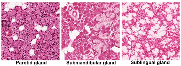
- Examine the images below and
sections of the
parotid gland, the
submandibular gland (sample
1, sample 2
 ),
and
sublingual gland. Compare
the relative amounts of serous and mucus secreting acini in
these glands. The parotid is purely serous secreting. Although
the submandibular and sublingual glands are mixed glands, the
sublingual gland contains relatively more mucous secreting acini.
Also, identify serous demilunes in the submandibular and
sublingual glands. ),
and
sublingual gland. Compare
the relative amounts of serous and mucus secreting acini in
these glands. The parotid is purely serous secreting. Although
the submandibular and sublingual glands are mixed glands, the
sublingual gland contains relatively more mucous secreting acini.
Also, identify serous demilunes in the submandibular and
sublingual glands.
-
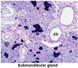 The PAS-alcian blue stain used
in this slide of
submandibular gland stains mucus (M) well, and shows
the distinct difference between the two types of secretory
cells. Look for striated ducts (SD) and larger
excretory ducts (ED) in the image at the right and these
slides (sample 1,
sample 2). Note
the tall columnar, pale staining epithelium of the striated
ducts and the stratified cuboidal to columnar epithelium lining
the excretory ducts. The PAS-alcian blue stain used
in this slide of
submandibular gland stains mucus (M) well, and shows
the distinct difference between the two types of secretory
cells. Look for striated ducts (SD) and larger
excretory ducts (ED) in the image at the right and these
slides (sample 1,
sample 2). Note
the tall columnar, pale staining epithelium of the striated
ducts and the stratified cuboidal to columnar epithelium lining
the excretory ducts.
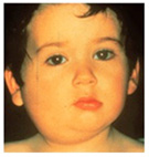 Clinical note: The childhood
disease called mumps is a viral infection of the salivary glands
(almost always the parotid glands), causing swelling and tenderness.
The disease is usually self-limiting but the virus can spread to
other organs, including the inner ear where it can lead to deafness. Clinical note: The childhood
disease called mumps is a viral infection of the salivary glands
(almost always the parotid glands), causing swelling and tenderness.
The disease is usually self-limiting but the virus can spread to
other organs, including the inner ear where it can lead to deafness.
Now for the esophagus. |