|
 Now that you are familiar with the
overall organization of the kidney, let’s study the structures of
the cortex in more detail. Now that you are familiar with the
overall organization of the kidney, let’s study the structures of
the cortex in more detail.
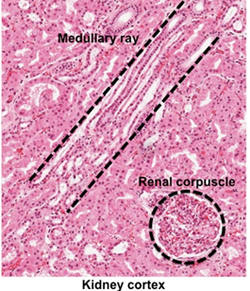
- Using these sections of human
kidney (sample 1,
sample 2
 ), examine the
structures in the cortex at higher magnification. Note the
presence of renal corpuscles and their
associated convoluted tubules, as well as the straight
tubules constituting the medullary rays, as seen in the
image at the right. ), examine the
structures in the cortex at higher magnification. Note the
presence of renal corpuscles and their
associated convoluted tubules, as well as the straight
tubules constituting the medullary rays, as seen in the
image at the right.
- Study figure 19-5 in Junqueira’s
Histology 14e and the image at the right of renal corpuscles.
Examine renal corpuscles on the slides at higher magnification,
and identify the simple squamous
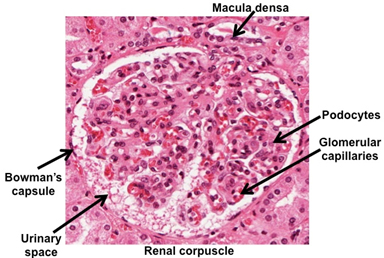 epithelium of
Bowman's
capsule, urinary space, glomeruli, and the large, pale podocytes.
Mesangial cells that have a variety of
functions (e.g. phagocytic and immune functions) are also
present among the podocytes, but they are difficult to identify. epithelium of
Bowman's
capsule, urinary space, glomeruli, and the large, pale podocytes.
Mesangial cells that have a variety of
functions (e.g. phagocytic and immune functions) are also
present among the podocytes, but they are difficult to identify.
- Try to find examples of renal
corpuscles that are cut to show the afferent or efferent
arterioles, macula densa, and juxtaglomerular apparatus.
It can be difficult to find these structures. The
juxtaglomerular apparatus contains specialized smooth muscle juxtaglomerular cells and
lacis cells (extraglomerular
mesangial cells). Juxtaglomerular cells secrete renin, a
protease that activates the renin-angiotensin-aldosterone system
involved in blood pressure regulation. The macula densa
regulates filtration rates in response to glomerular pressure
and tubular fluid composition. The extraglomerular mesangial
cells have functions similar to the mesangial cells associated
with the podocytes and glomeruli.
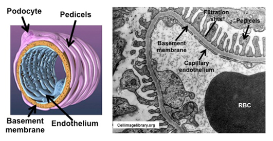
Study the diagram and electron
micrograph of glomeruli and their associated podocytes below and
identify:
- Podocytes and their pedicels
(foot processes)
- Fenestrated capillary
endothelial cells
- Glomerular basement membrane
- Urinary (Bowman's) space
Note the organization of the
podocyte pedicels and the endothelial cell fenestrations in
close association with their shared basement membrane. These
structures constitute the apparatus where blood is filtered to
produce the initial urine that collects in the urinary space
before it enters into the proximal convoluted tubule.
Clinical note: The
inflammatory condition known as glomerulonephritis results from
an autoimmune cross reaction on the part of the host against
streptococcal antigens. Infections triggering this reaction can
occur in either the skin or throat. The inflammatory response is
largely confined to the glomeruli, damaging the capillaries and
basement membrane to the extent that proteins and erythrocytes
may appear in the urine. The injury results from
antigen-antibody complexes getting stuck in the glomerular
filter.
Next
let's consider kidney infections and
look at examples of the nephron. |