|
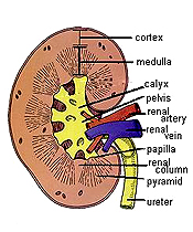 The kidney is a bean-shaped
organ with renal arteries and veins entering with the ureter at
the hilum. It consists of a cortex containing renal
corpuscles and their associated convoluted and straight
tubules and a medulla containing mainly straight tubules
and ducts. The renal corpuscles and their associated tubules
constitute the nephron, the basic functional unit of the
kidney involved in blood filtration and urine production. The kidney is a bean-shaped
organ with renal arteries and veins entering with the ureter at
the hilum. It consists of a cortex containing renal
corpuscles and their associated convoluted and straight
tubules and a medulla containing mainly straight tubules
and ducts. The renal corpuscles and their associated tubules
constitute the nephron, the basic functional unit of the
kidney involved in blood filtration and urine production.
In the medulla, the straight tubules
and ducts are organized into 8-15 renal pyramids. Each
renal pyramid is positioned with its base at the
corticomedullary junction and its tip (renal papillae) near the
renal pelvis in association with a minor calyx that leads into
the ureter. The pyramid is surrounded by cortical tissue (a
renal column of Bertin) that dips down into the medulla. The
renal pyramid with its associated cortical tissue constitutes a
renal lobe. Medullary tissue also extends up into the
cortex, forming medullary rays that are interspersed
between cortical tissue containing the renal corpuscles and
their associated convoluted tubules.
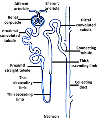
Study the diagrams at the right
showing the overall organization of the kidney and the
arrangement of structures within the nephrons. Refer to Figure
19-3 in Junqueira’s Histology 14e to review the blood supply to
the kidneys noting the relationships between the vasculature and
structures of the kidney. Click ere for a
larger, printable image.
Examine the overall histological
organization of the kidney, using this section of rat kidney
and the
image to the right. Unlike the human kidney, the rat kidney is unilobular, consisting of one large renal lobe with all of its
ducts converging in the direction of the hilum.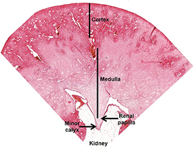
- Identify the
capsule and adipose-rich connective tissue at the hilum
surrounding the renal pelvis.
- Identify the cortex with its renal
corpuscles and the medulla with its straight tubules and
collecting ducts.
-
Near the corticomedullary junction, identify arcuate arteries and veins cut transversely.
- Identify the renal
papilla, noting that the largest collecting ducts (the ducts of
Bellini) converge here.
- Examine the calyx located near the renal
papilla. Note that the calyx is lined by transitional or urinary epithelium.
- Click here for a
large, printable image.
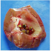
Clinical note: Kidney stones (nephrolithiasis) are
concretions of calcium salts and uric acid that can form in the
renal pelvis when urine contains high concentrations of
substances such as calcium and uric acid. Small stones may pass
down the ureters (with considerable pain) and larger stones can
be destroyed by focused sound waves in a procedure called
lithotripsy. (Photo courtesy of Dr. C.F. Verkoelen)
Now let's
take a look at the renal cortex. |