Human Structure Virtual Histology
|
Respiratory System, The Trachea |
|
|
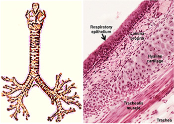 The
trachea conducts air to the two main bronchi, which are similar
to the trachea
histologically. Examine the diagram of trachea and major bronchi,
keeping in mind that as the trachea branches into the bronchi and
the bronchi branch into bronchioles, the components within the walls
(lining epithelium, hyaline cartilage, and smooth muscle) gradually
change. For example, when the trachea branches into the two main
bronchi, the C-shaped cartilage rings of the trachea give way to
irregular plates of hyaline cartilage in the bronchi. Progressive
changes to the walls of the airways will be a recurring theme as we
examine the lungs. The
trachea conducts air to the two main bronchi, which are similar
to the trachea
histologically. Examine the diagram of trachea and major bronchi,
keeping in mind that as the trachea branches into the bronchi and
the bronchi branch into bronchioles, the components within the walls
(lining epithelium, hyaline cartilage, and smooth muscle) gradually
change. For example, when the trachea branches into the two main
bronchi, the C-shaped cartilage rings of the trachea give way to
irregular plates of hyaline cartilage in the bronchi. Progressive
changes to the walls of the airways will be a recurring theme as we
examine the lungs.
-
 Examine this
longitudinal section of
larynx that leads into the trachea and this H&E stained
slides of trachea
(sample 1 Examine this
longitudinal section of
larynx that leads into the trachea and this H&E stained
slides of trachea
(sample 1  and
sample2). and
sample2).
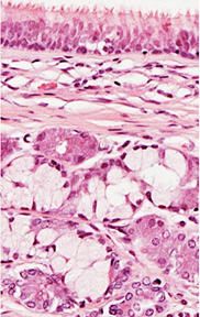
- Identify the layers in the wall
of the trachea, as seen in the slides and images above and at
the right, including the respiratory epithelium, noting
especially the cilia and thick basement membrane, the lamina
propria with serous-mucous glands, the hyaline cartilage, and
trachealis muscle.
-
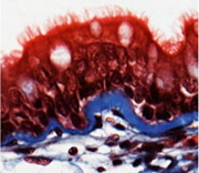 Study the epithelium in the
trichrome stained slide, noting the cilia, goblet cells,
as well
as the thick basement membrane that is nicely revealed by this
stain. Study the epithelium in the
trichrome stained slide, noting the cilia, goblet cells,
as well
as the thick basement membrane that is nicely revealed by this
stain.
On to the lung. |
|