Human Structure Virtual Histology
|
Respiratory System, The Larynx |
|
|
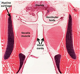 The
larynx, seen in the image at the right, is the beginning of the
lower respiratory tract and leads into the trachea. It works
together with the pharynx, tongue, and lips to allow for speech. The
epiglottis closes the larynx, preventing inspired air from entering
the esophagus and aspiration of food and liquids into the trachea.
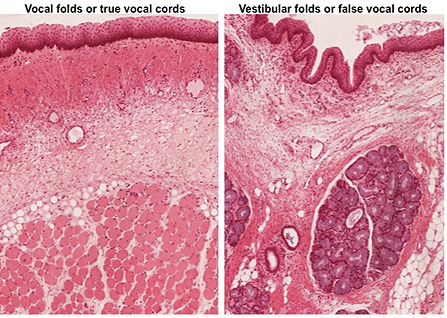
The larynx contains vocal folds or true vocal cords and vestibular
folds or false vocal cords that produce sounds and resonance,
respectively. Both are lined by stratified squamous epithelium and
in some areas, respiratory epithelium, with an underlying loose
connective tissue containing serous-mucous glands. The vocal folds
contain a thick layer of skeletal muscle (vocalis muscle), which
allows for movement of the folds to produce sounds. The
larynx, seen in the image at the right, is the beginning of the
lower respiratory tract and leads into the trachea. It works
together with the pharynx, tongue, and lips to allow for speech. The
epiglottis closes the larynx, preventing inspired air from entering
the esophagus and aspiration of food and liquids into the trachea.
The larynx contains vocal folds or true vocal cords and vestibular
folds or false vocal cords that produce sounds and resonance,
respectively. Both are lined by stratified squamous epithelium and
in some areas, respiratory epithelium, with an underlying loose
connective tissue containing serous-mucous glands. The vocal folds
contain a thick layer of skeletal muscle (vocalis muscle), which
allows for movement of the folds to produce sounds.
- Take a look at these samples of
larynx, an H&E stained
frontal section
of larynx and a trichrome stained, longitudinal section of
monkey larynx.
- Compare and contrast the features of
the vocal folds and vestibular folds in the slides and images below.
Note areas within the lining epithelium that change from stratified squamous epithelium to pseudostratified
columnar, ciliated epithelium (respiratory epithelium). Identify
the vocalis muscle in the vocal folds and serous-mucous glands
in the mucosa. Last, note the hyaline cartilage in the walls of
the larynx.
Now for the
trachea. |
|