 Veins
and Venules Veins
and VenulesVenules collect
blood from capillary networks and gradually merge to form veins to
carry blood from organs and
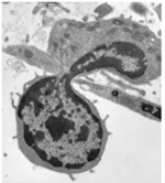
tissues back to the heart. Venules are also the main vessels from which immune cells
exit the circulation (i.e. undergo diapedesis) and enter the CT at
sites of injury or infection. Check out this TEM image to the right
where an immune cell can be seen squeezing between endothelial
cells.
-
 Examine examples of venules and
small veins in these three slides (sample
1, sample 2,
sample 3). Examine examples of venules and
small veins in these three slides (sample
1, sample 2,
sample 3).
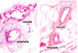
Compare the size, lumen shape, and wall thickness (relative
amounts of smooth muscle in the tunica media and CT in the
tunica adventitia) of venules and arterioles. Veins and arteries
often travel together and can be seen near each other in
histological specimens (as seen in the images to the right),
helping you to make these comparisons.
- Examine examples of medium size
veins in this
slide of CT
 .
Medium veins provide channels for blood flow from organs and
into large veins for return to the heart. Identify the 3 layers
or tunics seen earlier in arteries. The tunica media of medium
veins typically has 3-5 layers of smooth muscle, far fewer than
the 20-40 muscle layers found in medium arteries. .
Medium veins provide channels for blood flow from organs and
into large veins for return to the heart. Identify the 3 layers
or tunics seen earlier in arteries. The tunica media of medium
veins typically has 3-5 layers of smooth muscle, far fewer than
the 20-40 muscle layers found in medium arteries.
- Finally, study these sections of
large veins (sample 1 and
sample 2),
which function to return blood to the heart. Note how the
thickness of the wall and its various layers compare to the wall
of the neighboring artery. Large veins usually have 5 or more
layers of smooth muscle in their tunica media and are unique in
that they often have longitudinal bundles of smooth muscle in
their thick tunica adventitia.

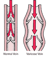
Veins contain valves to prevent
backflow of blood that’s flowing against gravity. These are
difficult to find in histological
specimens because they are thin and delicate, making it hard to
catch them in sections.
Clinical note: Weakness in the
walls or valves of veins can lead to abnormally dilated or varicose
veins, which most commonly occur in the lower legs where backflow of
blood is particularly common due to the pull of gravity.
The lymphatic
vessels are next. |