| Arterioles and Capillaries
The smallest branches of arteries are
called arterioles. The walls of arterioles are thin, the tunica
media has only 1-3 layers of smooth muscle, and the tunica
adventitia is a thin layer of CT. Arterioles control blood flow to
capillaries and resist blood flow, making them major regulators of
systemic blood pressure.
- Examine arterioles in the CT on
these slides (sample 1,
sample 2)
and in the image below. Compare the arterioles to their
companion venules and small veins, noting the irregular,
collapsed lumens of the veins and their relatively thinner
walls.

Capillaries usually have small
lumens, often no more than the diameter of erythrocytes. They are
composed of a lining endothelium and basement membrane, and lack the
typical tunics seen in other vessels. They function to exchange
oxygen, nutrients, and wastes with cells.
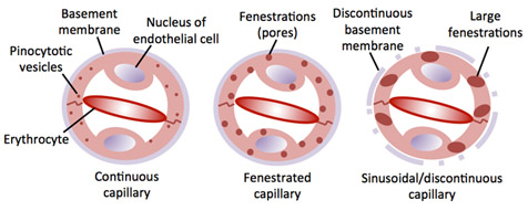
Capillaries can be classified
as one of three types:
- Continuous - endothelium
and basement membrane are continuous, lack fenestrations
(pores), and contain pinocytotic vesicles.
- Fenestrated - endothelium
contains pores.
- Sinusoidal - endothelium
contains very large pores and basement membrane may be
discontinuous.
-
 Examine
capillaries in this section of skeletal muscle
of the tongue
and CT of mesentery.
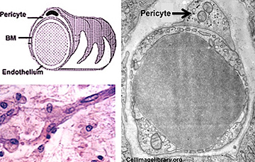
Also, look carefully for pericytes. These unique cells
modulate capillary functions and also serve as progenitor cells
to provide new cells for repairing damaged vessels. Examine
capillaries in this section of skeletal muscle
of the tongue
and CT of mesentery.
Also, look carefully for pericytes. These unique cells
modulate capillary functions and also serve as progenitor cells
to provide new cells for repairing damaged vessels.
-
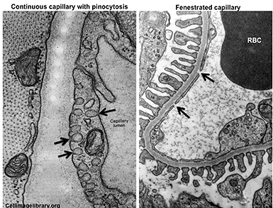 Finally, examine these electron
micrographs of the two common types of capillaries, noting the
pinocytotic vesicles often present in continuous capillaries and
the fenestrations seen in the very thin endothelium found within
capillaries of the kidney where blood is filtered to produce
urine. Finally, examine these electron
micrographs of the two common types of capillaries, noting the
pinocytotic vesicles often present in continuous capillaries and
the fenestrations seen in the very thin endothelium found within
capillaries of the kidney where blood is filtered to produce
urine.
Veins and
venules
|