|
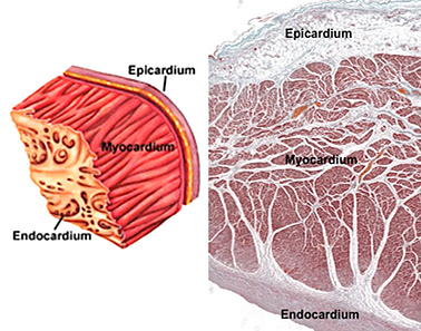 The Heart The Heart
Examine this
trichrome stained specimen and this
H&E stained specimen of the heart wall. The trichrome stained specimen nicely
demonstrates blood vessels and dense CT in the myocardium. Examine the major
features of each of the three layers of the heart on both slides and
in the images to the right.
of the heart wall. The trichrome stained specimen nicely
demonstrates blood vessels and dense CT in the myocardium. Examine the major
features of each of the three layers of the heart on both slides and
in the images to the right.
-
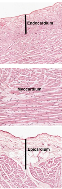 The
endocardium is lined by endothelium with underlying
layers of CT, a middle layer of smooth muscle and an elastic CT,
and a subendocardial layer that is connected to the myocardium.
The subendocardial layer contains modified cardiac muscle cells
(e.g.Purkinje fibers) of the heart’s impulse conducting
system. The thickness of the CT layers in the endocardium
varies. The
endocardium is lined by endothelium with underlying
layers of CT, a middle layer of smooth muscle and an elastic CT,
and a subendocardial layer that is connected to the myocardium.
The subendocardial layer contains modified cardiac muscle cells
(e.g.Purkinje fibers) of the heart’s impulse conducting
system. The thickness of the CT layers in the endocardium
varies.
- The myocardium is the
thickest layer of the heart wall and contains cardiac muscle,
connective tissue, and small blood vessels. It is also thicker in
the ventricles compared to the atria. Take a look at the image to
the right and note how the cardiac muscle fibers are cut in multiple
planes of section because these fibers are organized spirally around
each chamber.
- The epicardium is lined
by mesothelium with an underlying layer of loose CT that is rich
in adipose tissue and contains small nerves and blood vessels as
well as the large coronary vessels.
The myocardium
is next. |