|
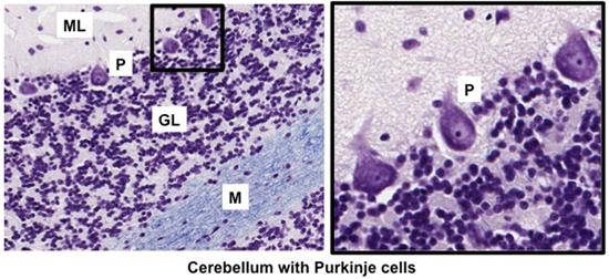
The cerebellum is organized as
three layers: outer molecular layer, thin middle layer of
Purkinje cells, and thick inner granular layer. The cerebellum
functions to coordinate the activities of muscles within the body.
Examine glial cells and other types of neurons in sections of the
cerebellum (sample 1 and
sample 2 and in the
images below. and
sample 2 and in the
images below. Identify:
- Neurons
of the molecular layer (ML)
- Neurons of the granular layer (GL)
- Purkinje
cells (P) (a type of neuron) between these layers
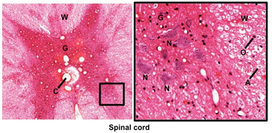
Examine these two
sections of spinal cord (sample 1 and
sample 2)
and the images below. In contrast to the cerebrum and cerebellum,
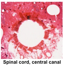
gray matter is internal to white matter in the spinal cord. Identify
the central gray matter (G), containing motor neurons (interneurons)
and abundant astrocytes, the central canal (C) lined by ependymal
cells (E), and the outer white matter (W), consisting largely of myelinated axons
(A) and oligodendrocytes (O). The myelin, composed
mainly of lipid, is lost during tissue preparation, leaving clear
circular spaces around the small eosinophilic axon when seen in
cross section.
Clinical note: The CNS is subject to various diseases
in which there is destruction of the myelin sheaths, such as
multiple sclerosis (MS). The functions and body regions affected by
MS depend on where in the CNS the focal areas of demyelination
occur. Weakness and paralysis of one or more limbs are common
sequelae of this disease.
Now for ganglia. |