|
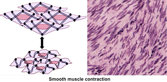
Smooth Muscle
This muscle type is found as layers
in the walls of most organs where it provides for involuntary
motility. Smooth muscle consists of layers or bundles of
non-striated, spindle-shaped fibers that contain one centrally
located nucleus per fiber. The actin and myosin filaments are
organized as a lattice-like network that is connected to the
sarcolemma via adhesion complexes called dense bodies. This
organization allows the cells to twist as they contract, sometimes
giving them a distinctive corkscrew appearance in specimens as
seen in the diagram and image below. In addition to lacking
striations, smooth muscle cells differ from striated muscle cells
because they also lack T-tubules.
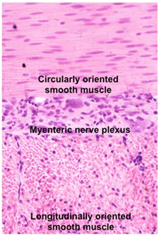 Examine smooth muscle cut
transversely and longitudinally in the
muscularis of the esophagus
and small intestine Examine smooth muscle cut
transversely and longitudinally in the
muscularis of the esophagus
and small intestine ,
and in the
myometrium of the uterus. Identify fibers, fascicles, and the
connective tissue layers, comparing these features to those of
skeletal muscle. The CT layers are generally not named in smooth
muscle. The muscularis of the gastrointestinal tract is frequently
organized as inner circular and outer longitudinal layers of smooth
muscle with a nerve plexus located in between the layers as seen in
the image at the right. ,
and in the
myometrium of the uterus. Identify fibers, fascicles, and the
connective tissue layers, comparing these features to those of
skeletal muscle. The CT layers are generally not named in smooth
muscle. The muscularis of the gastrointestinal tract is frequently
organized as inner circular and outer longitudinal layers of smooth
muscle with a nerve plexus located in between the layers as seen in
the image at the right.
Now try this self-assessment
quiz, no one sees the score but you.
On to skin. |