|
Monocytes and platelets
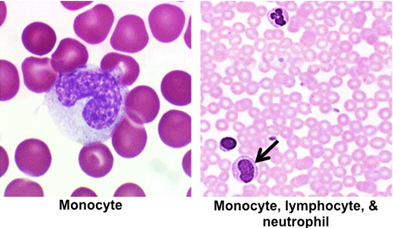
Monocytes are agranulocytes that are
produced in the bone marrow. These cells travel to organs of the
body where they enter into the connective tissue and differentiate
into phagocytic and antigen presenting cells, such as macrophages,
dendritic cells, and osteoclasts. These cells have major roles in
clearing apoptotic cells, removing debris, ingesting pathogens, and
mediating immune responses.
- Examine the images below and the
peripheral blood smear
again.
Identify monocytes, noting their size (12-15 microns in diameter),
pale basophilic cytoplasm, and their large, indented or C shaped
nuclei.
- To review the ultrastructural
features of monocytes and platelets in TEM images, please see
Figures 12-12 and 12-13 in Junqueira’s Histology 14e
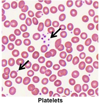 Platelets (thrombocytes) are small, round to oval cellular
fragments (2-4 microns) that normally range in number between
150,000-400,000 per microliter of blood. Platelets are essential for
blood stasis and healing of damaged blood vessels. They contain cell
surface proteins that allow them to adhere to collagen exposed by
wounds in the vasculature, helping to plug the wound. They also
possess a variety of granules with numerous substances involved in
blood clotting, clot retraction, and clot removal. Platelets are
produced in the bone marrow as cellular fragments that are released
from megakaryocytes. Megakaryocytes are not present in peripheral
blood smears. We will look at megakaryocytes when we examine a bone
marrow smear in a moment. Platelets (thrombocytes) are small, round to oval cellular
fragments (2-4 microns) that normally range in number between
150,000-400,000 per microliter of blood. Platelets are essential for
blood stasis and healing of damaged blood vessels. They contain cell
surface proteins that allow them to adhere to collagen exposed by
wounds in the vasculature, helping to plug the wound. They also
possess a variety of granules with numerous substances involved in
blood clotting, clot retraction, and clot removal. Platelets are
produced in the bone marrow as cellular fragments that are released
from megakaryocytes. Megakaryocytes are not present in peripheral
blood smears. We will look at megakaryocytes when we examine a bone
marrow smear in a moment.
- Identify platelets in the peripheral blood
smear. Note their small size, lack of nuclei, distribution (singly
or in clusters), and basophilia relative to the RBCs.
So, where does
it all happen? |