|
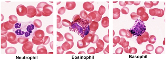
Granulocytes
Granulocytes, as their name implies,
contain distinctive cytoplasmic granules that are easily visualized
in peripheral blood smears stained with Wright’s stain. There are
three types of granulocytes: neutrophils, eosinophils, and
basophils. These cells are approximately 12-15 microns in
diameter, and their nuclei are either segmented or bilobed. In the
case of eosinophils and basophils, their nuclei may be obscured by
their abundant, intensely stained specific granules.
Neutrophils are the most abundant
granulocyte, representing 50-70% of the WBC population, and are
therefore the most readily identifiable leukocyte in a peripheral
blood smear. They contain many lysosomes or azurophilic granules and
pale staining specific granules, making it easy to visualize their
segmented nuclei. Neutrophils are among the first cells to arrive at
the site of injury or infection, exiting the vasculature by the
process of diapedesis. They enter into the connective tissue where
they function as phagocytes, engulfing and killing bacteria and
releasing cytokines that modulate inflammation.
Eosinophils represent about 1-4%
of white blood cells in a peripheral smear and are more difficult to
locate. They possess distinctive, intensely stained pink granules
that may obscure their bilobed nucleus. Eosinophils are involved in
immune responses to helminthic infections and allergic reactions.
Basophils are the least abundant
granulocyte, representing 0.5-1% of the WBC count. They possess
abundant, basophilic specific granules and a bilobed or sometimes S
shaped nucleus. They possess cell surface receptors for IgE, and
binding to IgE triggers secretion of the contents in their specific
granules. Basophils modulate immune responses and are involved in
allergic and hypersensitivity reactions.
- Use the images below to help you
identify the granulocytes in these slides of peripheral blood
smears (sample 1
and sample 2).
Note their size compared to red blood cells, nuclear morphology,
and the appearance of their granules.
- To review the ultrastructural
features of granulocytes in TEM images, please see Figures 12-8
through 12-10 in Junqueira’s Histology 14e.
Clinical note: A large
accumulation of neutrophils, especially dead and dying
neutrophils that have engulfed many bacteria, constitutes “pus.”
Pus formation is a normal part of an infection that occurs
within tissues.
Want to try for
a lymphocyte? |