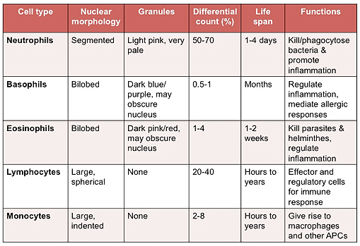
| Review the table below describing
the characteristics and functions of mature white blood cells (WBCs
or leukocytes). It is useful to keep in mind the normal differential
counts of WBCs in the circulation for a variety of reasons. When
looking at blood smears, this knowledge will help you gage how often
you might expect to find the various cell types (e.g. neutrophils
and lymphocytes are relatively easy to find, while basophils are
much harder to locate).

Examine a human
peripheral blood
smear, containing mature red blood cells (RBCs,
erythrocytes) and leukocytes. This smear was prepared with
polychromatic Wright's stain, which stains both basophilic and
acidophilic cellular components. containing mature red blood cells (RBCs,
erythrocytes) and leukocytes. This smear was prepared with
polychromatic Wright's stain, which stains both basophilic and
acidophilic cellular components.
-
 Identify the red blood cells,
which are the most numerous cells in the smear as they represent
~37-52% of the formed elements in blood. RBCs have a uniform
diameter of ~7 um and can be found in the capillaries of most
tissues. Their size provides a “histological ruler” by which to
estimate sizes of other cells and tissue structures. Identify the red blood cells,
which are the most numerous cells in the smear as they represent
~37-52% of the formed elements in blood. RBCs have a uniform
diameter of ~7 um and can be found in the capillaries of most
tissues. Their size provides a “histological ruler” by which to
estimate sizes of other cells and tissue structures.
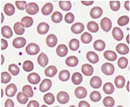
- Study the RBCs closely in the
slide and image at the right. Note the intense eosinophilia and
lack of basophilia within the cytoplasm due to the presence of
abundant hemoglobin and absence of rER, respectively. Also,
notice the central pallor of the RBC. This feature results from
the RBC’s unique biconcave shape that facilitates gas exchange.
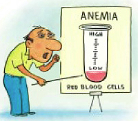
 Clinical
note: Anemia is the term applied to any significant reduction in
the total mass of erythrocytes or in their content of hemoglobin.
Iron-deficiency anemia results from having too little iron available
for hemoglobin synthesis. The hereditary disease sickle-cell anemia
involves a mutation that produces a single amino acid substitution
in hemoglobin, which makes the protein crystallize within
erythrocytes at low-oxygen tensions. This alters the cells overall
shape and can lead to microvascular blockage and various other
problems. Clinical
note: Anemia is the term applied to any significant reduction in
the total mass of erythrocytes or in their content of hemoglobin.
Iron-deficiency anemia results from having too little iron available
for hemoglobin synthesis. The hereditary disease sickle-cell anemia
involves a mutation that produces a single amino acid substitution
in hemoglobin, which makes the protein crystallize within
erythrocytes at low-oxygen tensions. This alters the cells overall
shape and can lead to microvascular blockage and various other
problems.
Now let's take a look at some
white blood cells. |