|
 Study
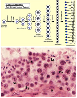
the schematic diagram of sperm development and the image below,
noting the relationships of the developing sperm and sustentacular
Sertoli cells. Next, examine these two examples of testis (sample1,
sample 2 Study
the schematic diagram of sperm development and the image below,
noting the relationships of the developing sperm and sustentacular
Sertoli cells. Next, examine these two examples of testis (sample1,
sample 2 at
higher magnification. Carefully examine the cells inside a seminiferous tubule and distinguish sperm in the various stages of
spermatogenesis and spermiogenesis. In a tubule cut transversely,
identify: at
higher magnification. Carefully examine the cells inside a seminiferous tubule and distinguish sperm in the various stages of
spermatogenesis and spermiogenesis. In a tubule cut transversely,
identify:
- Sustentacular or Sertoli
cells (Sc) (with their distinctive nucleoli)
- Spermatogonia (Sg)
- Primary spermatocytes (Ps)
- Secondary spermatocytes are
short-lived and difficult to find, so dont spend time looking
for these
- Early and late spermatids
(Es, Ls)
- Spermatozoa (Sz)
- Myoid cells (M)
 Finally, in the interstitial
tissue between the seminiferous tubules, identify the
interstitial cells or Leydig cells as seen in the image at
the right. These cells are large, round pale-staining cells.
They secrete testosterone, which is taken up by the Sertoli
cells and concentrated within the seminiferous tubules where it
serves an essential role in spermatogenesis. Finally, in the interstitial
tissue between the seminiferous tubules, identify the
interstitial cells or Leydig cells as seen in the image at
the right. These cells are large, round pale-staining cells.
They secrete testosterone, which is taken up by the Sertoli
cells and concentrated within the seminiferous tubules where it
serves an essential role in spermatogenesis.
Clinical notes: Failure of the
testes to descend into the scrotum during fetal development (cryptorchidism)
maintains their temperature at 37℃. This temperature inhibits
spermatogenesis, although testosterone production can still occur.
Excessive blood flow or dilated vasculature in the scrotum (varicocoele)
is another potential cause of infertility and can be surgically
corrected.
Most cases of testicular cancer (95%)
are of germ cell origin. The rest are derived from Sertoli cells or
Leydig cells. IUSMs own Dr. Lawrence Einhorn discovered a cure for
testicular cancer over 40 years ago.
Now for the
intratesticular ducts. |