|
Liver
-- is responsible for many different functions and is unusual in
being supplied by both arterial blood (for oxygen) and venous blood
(with nutrients for processing).
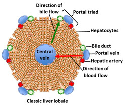 Review the structural organization of
the liver parenchyma, noting inflow of blood to
lobules from branches of both the hepatic artery and hepatic portal
vein and outflow through central venules to branches of the hepatic
vein. Note also how bile originates within lobules and is drained
via branches of the bile duct. A simplified version of one liver
lobule in transverse section is shown in the diagram to the right.
Click here for a larger view. Review the structural organization of
the liver parenchyma, noting inflow of blood to
lobules from branches of both the hepatic artery and hepatic portal
vein and outflow through central venules to branches of the hepatic
vein. Note also how bile originates within lobules and is drained
via branches of the bile duct. A simplified version of one liver
lobule in transverse section is shown in the diagram to the right.
Click here for a larger view.
List
several unrelated functions of hepatocytes.
 Clinical note: Hepatitis
involves infection or inflammation of hepatocytes and other
epithelial components in the liver. Cirrhosis involves excessive
proliferation of fibroblasts and collagen deposition in the stroma
of the liver. Hepatocytes have a remarkable capacity for
regeneration, but chronic, long-term alcoholism leads to the death
of these cells and cirrhosis. Clinical note: Hepatitis
involves infection or inflammation of hepatocytes and other
epithelial components in the liver. Cirrhosis involves excessive
proliferation of fibroblasts and collagen deposition in the stroma
of the liver. Hepatocytes have a remarkable capacity for
regeneration, but chronic, long-term alcoholism leads to the death
of these cells and cirrhosis.
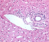 Examine
these sections of liver (sample
1, sample 2,
sample 3)
with the low
magnification, locate a set of portal tracts outlining a lobule. With the higher power objectives, identify the
central venule
in the center of the lobule and the "triad" of
components in a portal tract, namely branches of the hepatic portal
vein, hepatic artery, and bile duct. Examine
these sections of liver (sample
1, sample 2,
sample 3)
with the low
magnification, locate a set of portal tracts outlining a lobule. With the higher power objectives, identify the
central venule
in the center of the lobule and the "triad" of
components in a portal tract, namely branches of the hepatic portal
vein, hepatic artery, and bile duct.
What do the 3 components of a
“portal triad’ transport?
What fourth structure is also
usually there?
Detail of the
hepatic lobule. |