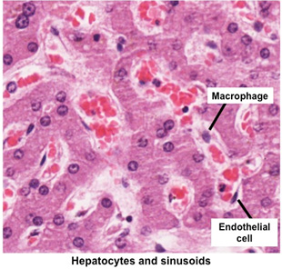
| Now, let’s take a closer look at the
hepatocytes and sinusoids. The sinusoidal arrangement of the
capillaries running between the cords of hepatocytes facilitates
rapid exchange of nutrients, metabolites, and endocrine products
between the hepatocytes and blood.
  Examine these two slides of liver (sample
1 H&E, and sample 2
Masson's trichrome) and the image below. Examine these two slides of liver (sample
1 H&E, and sample 2
Masson's trichrome) and the image below.
- Note the anastomosing
plates of hepatocytes between the portal tracts and the central
venule.
- Identify the vascular sinusoids between the plates of hepatocytes and the endothelial cells lining the sinusoids.
- Identify
the scattered, rounded macrophages (Kupffer cells) in the sinusoidal
lining.
- Note how the hepatocytes near the portal tract appear
different from those near the central vein. This is due to exposure
of the hepatocytes to blood with changing levels of oxygen and
metabolites as it flows from the portal areas towards the central
venule.
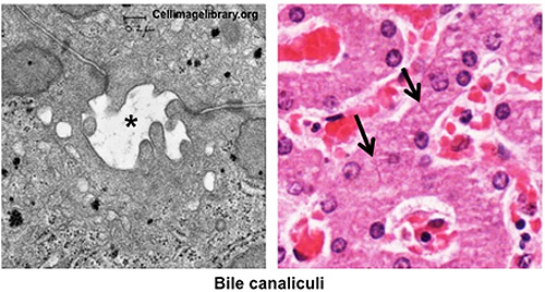
Next, carefully examine the ultrastructure of hepatocytes
forming a bile canaliculus (asterisk) in the electron micrograph and
light micrograph (arrows) images below. Bile is the major exocrine
product of the liver. It flows from hepatocytes into the bile
canaliculi, into the bile ductules at the portal areas, into the
bile ducts, and eventually into the common hepatic duct.
Finally,
study figure 16-14 from Junqueira’s Histology 14e to see additional
views of important ultrastructural features of the liver that you
should be familiar with, including:
- Microvilli projecting from the
hepatocytes
- Sinusoids
- The perisinusoidal space (of Disse)
- The bile
canaliculi
Now for the
gallbladder. |