|
 This
class of primary tissue supports and physically connects
other tissues and cells to form functional organs. Its fluid
provides a medium through which cells receive metabolic
support, such as diffusion of both nutrients and waste
products. This
class of primary tissue supports and physically connects
other tissues and cells to form functional organs. Its fluid
provides a medium through which cells receive metabolic
support, such as diffusion of both nutrients and waste
products. Generally,
tissues can be said to consist of both cells and an extracellular
matrix (ECM). ECM is composed of both protein fibers (collagen,
elastic, reticular fibers) and ground substance (glycosaminoglycans
[GAGs], proteoglycans, and multiadhesive glycoproteins). In
connective tissue (CT), the ECM component predominates relative to
the cellular component. In other words, cells tend to be widely
separated among ECM fibers and ground substances.
Functions of CT include the following:
- Structure, support, and
mechanical protection: capsules and fibrous septa of organs,
support and space filling (padding) of organs and tissue
elements, provides resistance to stress and shearing forces
- Nutrition: absorption in
gastrointestinal tract and energy storage in adipose tissue
- Defense: immune cells in
CT are a first-line of defense against microorganisms that
breach an epithelium
- Repair: scar formation
during wound healing
- Transport: continuous
formation and flow of interstitial fluid
Various combinations of cells,
fibers, and ECM components — both in numbers and types — create
different types, or classifications, of CT; however, the lines
separating these classifications are sometimes not distinct but
graded. We can classify different types of the CT into three
broad categories: embryonic, CT proper (loose and dense), and
specialized CT. We can further classify CT in the following way:
Embryonic CT
- Mesenchyme
- Mucous CT (Wharton’s jelly)
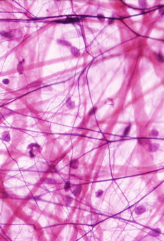
Loose CT Proper
- Areolar (seen in the
image on this page)
- Reticular
Dense CT Proper
- Dense irregular CT
- Dense regular CT
Specialized
Connective Tissue
- Cartilage:
hyaline, elastic, fibrocartilage
- Tendons and
ligaments
- Bone: spongy and
compact
- Blood
- Adipose tissue:
white and brown (Of the specialized CTs, only
adipose tissue will be described in this module;
the others are discussed in other modules)
The learning
objectives for this module are:
- Recognize
the major types of connective tissue proper
in light micrographs.
- Explain how
the structure of each major type of
connective tissue reflects its function.
- Distinguish
the various cells found in connective tissue
(fibroblasts, adipocytes, mast cells, plasma
cells, macrophages, and undifferentiated
mesenchymal cells), and describe their
functions and key features.
- Recognize
variations in distribution of ground
substance and the three types of
extracellular fibers in connective tissue
proper.
- Recognize
the unique features of adipose tissue,
comparing and contrasting these in white and
brown adipose tissue.
We will start
with embryonic (primitive)
connective tissue. |