|
 The
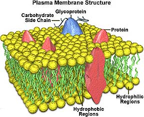
plasma membrane, or plasmalemma, of a cell consists of a
phospholipid bilayer with associated integral and peripheral
proteins, glycoproteins and glycolipids, and cholesterol. The plasma
membrane is only 7–10 nm thick, which is below the resolution of the
light microscope. The
plasma membrane, or plasmalemma, of a cell consists of a
phospholipid bilayer with associated integral and peripheral
proteins, glycoproteins and glycolipids, and cholesterol. The plasma
membrane is only 7–10 nm thick, which is below the resolution of the
light microscope.
The plasma membrane has several major
functions and serves to:
- Provide a physical barrier that
protects, supports, and separates the cytoplasm from the
extracellular environment
- Regulate the traffic of ions,
gases, nutrients, and wastes in and out of the cell
- Establish and maintain
electrochemical gradients between the intracellular and
extracellular environment
- Provide attachment and
communication with other cells and the extracellular matrix
- Participate in cell signaling
events that direct cell growth, division, survival, motility,
and differentiation
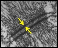 In very high
magnification, osmium-fixed TEM specimens, the structure of the
plasma membrane can be seen as a dark or electron dense line. This
specimen shows a desmosome where two adjacent plasma membranes
connect the intermediate filament network of these neighboring
cells. One of these plasma membranes can be seen between the tips of
the arrows. In very high
magnification, osmium-fixed TEM specimens, the structure of the
plasma membrane can be seen as a dark or electron dense line. This
specimen shows a desmosome where two adjacent plasma membranes
connect the intermediate filament network of these neighboring
cells. One of these plasma membranes can be seen between the tips of
the arrows.
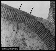 A thick external coat of
glycoproteins and glycolipids on the outside of the plasma membrane
is called a glycocalyx. Many cells have a glycocalyx, but it is
especially well developed on the microvilli of absorptive cells
lining the small intestine (arrows). A thick external coat of
glycoproteins and glycolipids on the outside of the plasma membrane
is called a glycocalyx. Many cells have a glycocalyx, but it is
especially well developed on the microvilli of absorptive cells
lining the small intestine (arrows).
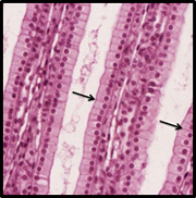 Although the plasma membrane is
generally not visible in the light microscope, occasionally its
presence can be discerned, such as in the epithelial cells lining
the
collecting ducts of the kidney medulla,
due to an abundance of associated proteins that stain with eosin
(arrows). Although the plasma membrane is
generally not visible in the light microscope, occasionally its
presence can be discerned, such as in the epithelial cells lining
the
collecting ducts of the kidney medulla,
due to an abundance of associated proteins that stain with eosin
(arrows).
Let's now take a look at
membrane
bound organelles within the cell. |