| The artifacts that can cause
confusion are due to problems or imperfections in the technique of
slide preparation and must not be interpreted as structural features
of the tissue shown. The
following artifacts are common and may be present in your slide
collection. Wrinkles, folds, and knife marks are especially hard to
avoid completely and you should expect these artifacts as they are
encountered.
- Wrinkles or folds: well-defined
dense-staining regions in the section where detail is obscured.
- Knife marks: straight cuts or
fine lines across the section caused by nicks in the microtome
knife.
- Poor fixation (postmortem
degeneration): the tissue stains poorly and shows poor
microscopic
detail.
- Shrinkage or small tears:
components are separated from each other giving rise to empty
spaces.
- Precipitates: usually appear as
small black particles on the section.
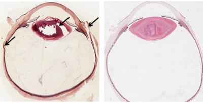
In the accompanying images
the arrows indicate knife marks, folds and tears in the eye specimen
on the left compared to the specimen on the right. Also, note the
variability of colors and staining intensity that can occur with H&E
staining.
Cell structures
and organelles. |