Much goes into making the slides you will
be studying in this course.
-
 Many
samples are of human origin and come from autopsy or surgical
procedures. In all cases, permission was given by the patient or
family to use the tissue for teaching purposes. Many
samples are of human origin and come from autopsy or surgical
procedures. In all cases, permission was given by the patient or
family to use the tissue for teaching purposes.
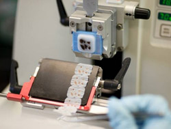
- In this exercise, we will first
look at the method of glass slide preparation so that you will
have the understanding of where the scanned slides came from.
- This unit will also include a
short segment on use of the optical as well as virtual
microscope.
Objectives for this laboratory:
- Describe the histological
techniques used in preparing samples for light and electron
microscopy.
- Explain the mechanism of
biological staining (acidophilia and basophilia) and the way in
which common stains are used to provide information about cells
in light microscopy.
- Interpret images of cells,
tissues, and organs when seen with both the light and electron
microscopes.
Have you
ever wondered where the tissues come from and how
the slides are made? |