|
Nerve tissue is a complex
organization of neurons, glial cells, vasculature, and connective
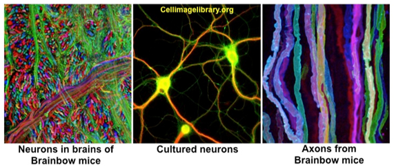
tissue coverings. Neurons are specialized to conduct impulses in
response to stimuli or environmental signals. To carry out this
function, neurons have distinctive morphologies (as seen in the
striking images below), possessing long cytoplasmic extensions
(axons and dendrites), which allow them to form hundreds of
connections with other neurons and cell types. Neurons are supported
by glial cells, which are involved in nutrition, protection, and the
formation of insulating sheaths.
Nerve tissue forms a complex,
interconnected communication system throughout the body and is
generally organized into two major divisions: the central nervous
system (CNS, includes the brain and spinal cord) and peripheral
nervous system (PNS, includes cranial, spinal, and peripheral nerves
and ganglia located outside of the CNS). The PNS and CNS work
together to receive, interpret, and integrate information in order
to regulate a diverse range of essential physiological (e.g.
respiration and blood pressure) and behavioral (e.g. defense and
feeding) processes.
The purpose of this unit is to
familiarize you with the histological features and functions of
nerve tissue. It is not necessary for you to identify distinct
functional regions within the central nervous system. The structures
of the CNS will be covered in detail in the Neuroscience and
Behavior course next semester.
The learning objectives for this unit
are:
- Identify neurons and distinguish
them from glial cells (astrocytes, oligodendrocytes, and
ependymal cells) in sections of spinal cord and brain using
light microscopy.
- Recognize neuronal cell bodies
and satellite cells in sections of ganglia, and explain their
functions.
- Identify Schwann cells, nodes of
Ranvier, fibroblasts, and connective tissue layers in sections
of peripheral nerves.
Slides and
Micrographs |