|
Compact and Trabecular Bone
Unlike the ground bone specimens, the
specimens of two small bones on the following slides were
decalcified chemically and then mounted and sectioned with a
microtome. After staining with H&E all of the cells are readily
distinguishable.
- Examine these three specimens
and the images below to compare compact and trabecular bone.
Bone sample 1 and
sample 2
show osteons very well.
Sample 3
 is mostly trabecular bone, but a small area of
compact bone can also be seen. Compare the appearance of the
osteons to those you saw in
the ground specimen.
Identify periosteum, endosteum, Haversian canals, osteocytes,
and marrow. is mostly trabecular bone, but a small area of
compact bone can also be seen. Compare the appearance of the
osteons to those you saw in
the ground specimen.
Identify periosteum, endosteum, Haversian canals, osteocytes,
and marrow.

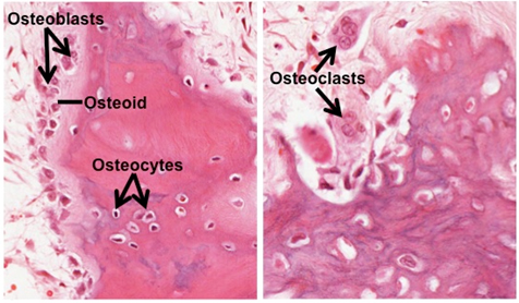
- The rest of the bone tissue
present on sample 3 is trabecular bone, and one area of this
specimen was undergoing fracture repair. There is much to
identify and study on this slide. Locate and carefully study the
following structures: periosteum, marrow, osteoblasts and
osteoid, osteoclasts, which may be in Howship’s lacunae, and
endosteum. Study the image below to help you find some of these
structures.
 Ossification
and bone formation. |