The pancreas is a dual
exocrine and endocrine gland. The exocrine pancreas is specialized
for secretion of digestive enzymes (e.g. proteinases, lipases,
amylases, and nucleases) that are carried by a duct system to the
duodenum. Small areas of endocrine tissue (the islets of Langerhans)
are interspersed amongst the exocrine pancreas and secrete key
hormones (insulin, glucagon, and somatostatin) that control blood
glucose levels.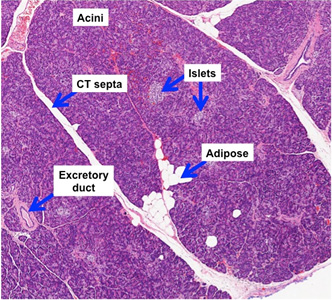
Examine sections of pancreas (sample
1 , sample 2,
sample 3)
with low power and observe the overall organization. , sample 2,
sample 3)
with low power and observe the overall organization.
- Lobules separated by CT septa
- Adipocytes
- Pale-staining pancreatic islets
of Langerhans
- Densely packed secretory acini
- Excretory ducts
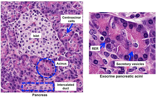
Using
higher magnification, examine
sample
1
in more detail. Identify acini in an area where the polarized nature
of the cells is apparent.
- Note that the lumens of
pancreatic acini are almost too small to see with the light
microscope.
- When examining the acinar cells,
note the basophilic staining in the basal region due to abundant
RER and the eosinophilic staining in the apical region due to
abundant protein-rich secretory vesicles.
- Examine the pale staining
centroacinar cells at the center of the acini leading into the
intercalated ducts lined by pale staining, low cuboidal
epithelium.
- Examine the ducts, recalling
that the intercalated ducts lead into intralobar, interlobular,
and then into the main pancreatic duct. The ductal epithelium
transitions to simple columnar and eventually to stratified
cuboidal-columnar epithelium in the larger excretory ducts.
Pancreatic islets (of Langerhans). |