Human Structure Virtual Histology
| Use of the light microscope, the
condenser and iris adjustment. |
|
|
|
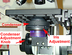 |
Every now and then,
someone will have fiddled with the position of the condenser.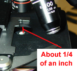
- If it's too high, your slide will have a real
grainy look and you'll never be able bring the image into focus.
- The top of the condenser should be about 1/4
of an inch below the slide you're viewing.
- Use the condenser adjustment knob to get it
where it belongs.
|
The diaphragm of the iris is the
mechanism that directs the light in a straight-line path through the
slide.
The
iris adjustment also seems to be a favorite for people to monkey
with.
- If it's too wide open, it might be
difficult to get the image into sharp focus.
- If it's to tight, the field will be
dim and the cells and fibers will have an annoying refractile
quality. Although sometimes this effect can be helpful. An example
is looking at RBCs that contain sickled hemoglobin.
- To adjust, move the little iris
lever (a little flat lever with a serrated edge) all the way to the
right and then open it back up to about 1/4 of the way.
- If you are looking in the oculars
while doing this, you will see the field lighten up and the
refractile quality of the fiber structures disappear.
Sum it up please and
tell me about this virtual microscope. |
|
Lab Table of Contents
| Glossary
Copyright 2016,
the Trustees of Indiana University
|