| Simple columnar
Simple columnar epithelial cells with
cilia are numerous in the lining of the oviduct, where they are
interspersed with secretory cells. where they are
interspersed with secretory cells.
- Compare the appearance of the
cilia in this epithelium with the striated border (microvilli)
of absorptive epithelium on the last slide.
- Examine the ultrastructural
views of the cilia and
microvilli shown in cross section and in
longitudinal section. Compare cilia (below) to the absorptive stereocilia at the apical surface of the pseudostratified
columnar cells lining the epididymis (epididymis 1, and
epididymis 2, and seen below).
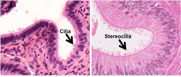
- Cilia have a core of
microtubules with associated motor proteins that allow them to
beat, which in the oviduct facilitates movement of the ovum
toward the uterus. In contrast, microvilli and stereocilia have
a core of actin microfilaments and function to increase surface
area for absorption.
- Learning about this is not just
an academic exercise; rather, it has real clinical significance.
Kartagener’s disease is an
inherited disorder involving mutations in the gene for dynein or
one of the many other proteins in cilia. All cilia in such
individuals are immotile, leading to infertility and chronic
respiratory disorders.
How about
pseudostratified and stratified epithelium? |