Epithelium, one of the four basic
tissue types of the body, is characterized by:
- An apical or free surface
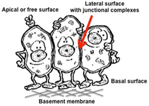
- A basal lamina (basement
membrane), which anchors the basal surface to underlying
structures
- Polarity due to distinct lipid
and protein components at each surface and localization of
organelles within the cytoplasm
- Close apposition and adhesion to
neighboring epithelial cells
In general, epithelia function to:
- Line and protect the surfaces of
the body and organ cavities (e.g. oral mucosa and lining
epithelium of the esophagus)
- Allow for absorption (e.g.
nutrients and water in the gastrointestinal tract)
- Produce
secretions (e.g. mucous, digestive enzymes, and hormones)
The classification of an epithelial
layer is based on three features:
- The shape of cells that comprise
its free surface (squamous, cuboidal, or columnar)
- The number of cellular layers
(one layer = simple, more than one layer = stratified)
- Specializations present at the
apical or free surface (keratinization, cilia, microvilli, or
stereocilia)
The learning objectives for this
module are:
- Classify epithelia by their
major structural types and recognize these using light
microscopy.
- Recognize by light microscopy
the basic morphological features and staining differences within
the secretory elements of serous and mucous exocrine glands.
Let's start with
simple squamous epithelium |